. 2015 Apr 2;16(4):400-12.
doi: 10.1016/j.stem.2015.02.006.

Basal cell carcinoma preferentially arises from stem cells within hair follicle and mechanosensory niches

Affiliations

Basal cell carcinoma preferentially arises from stem cells within hair follicle and mechanosensory niches

Shelby C Peterson et al. Cell Stem Cell. .

Abstract

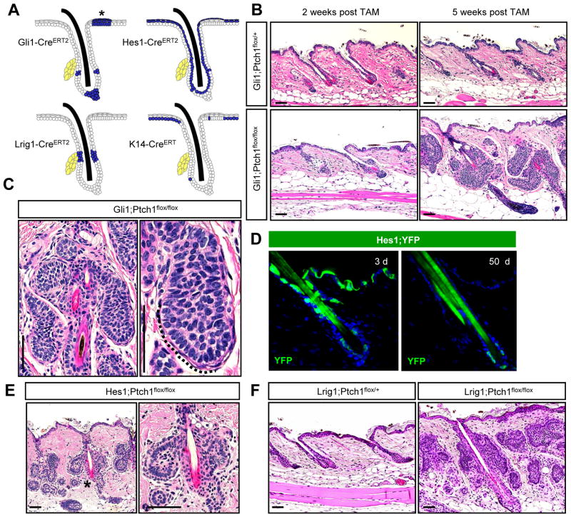
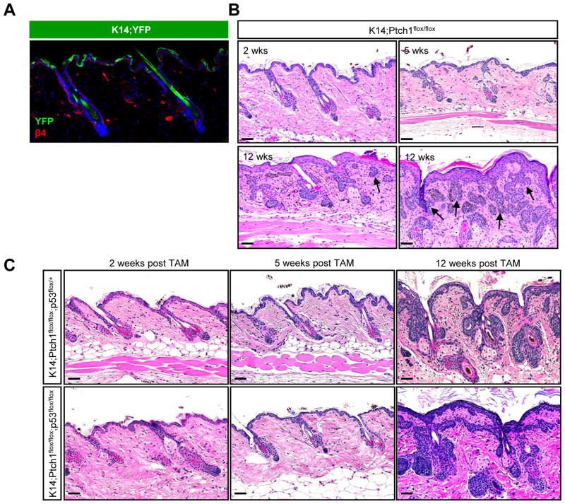
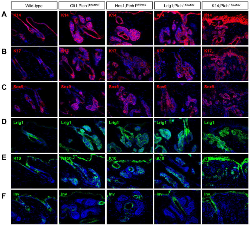
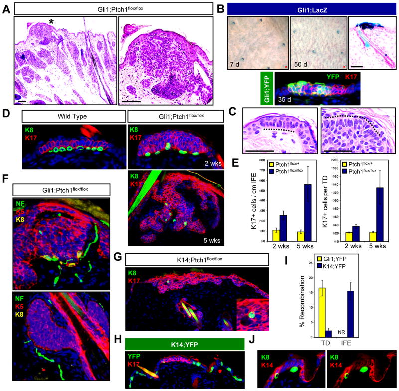
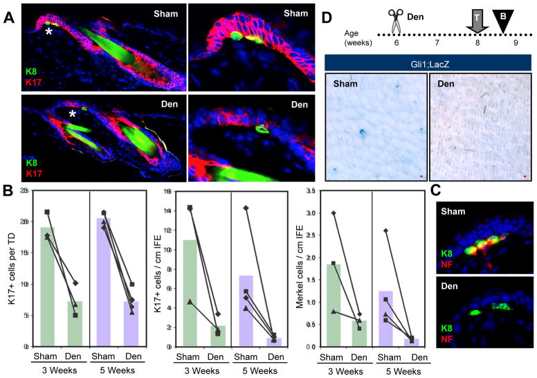
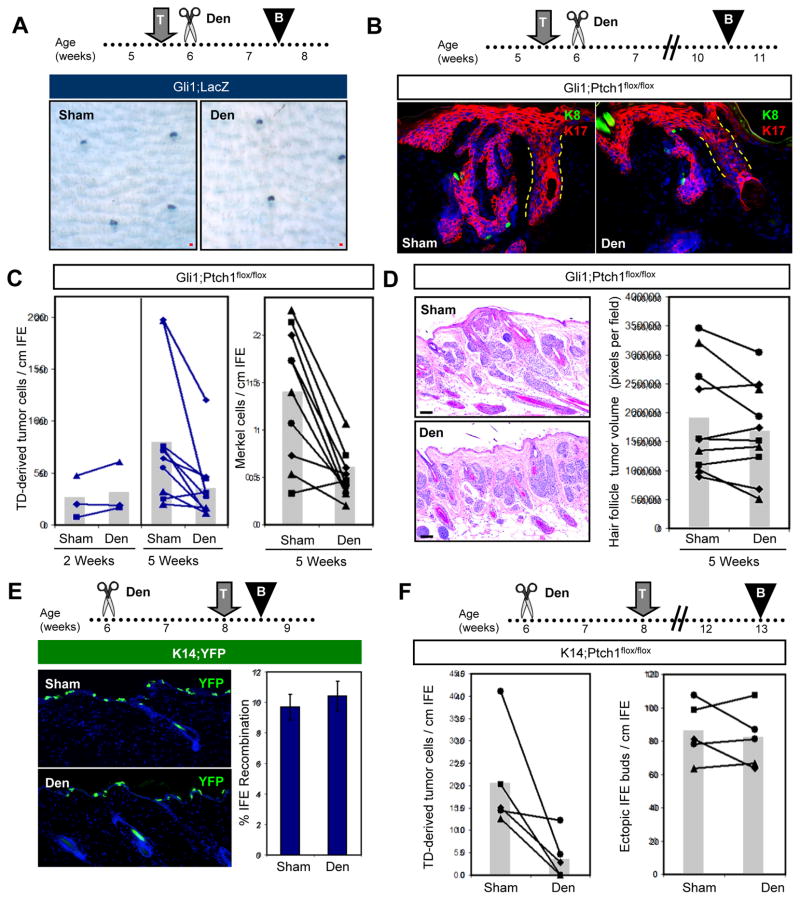
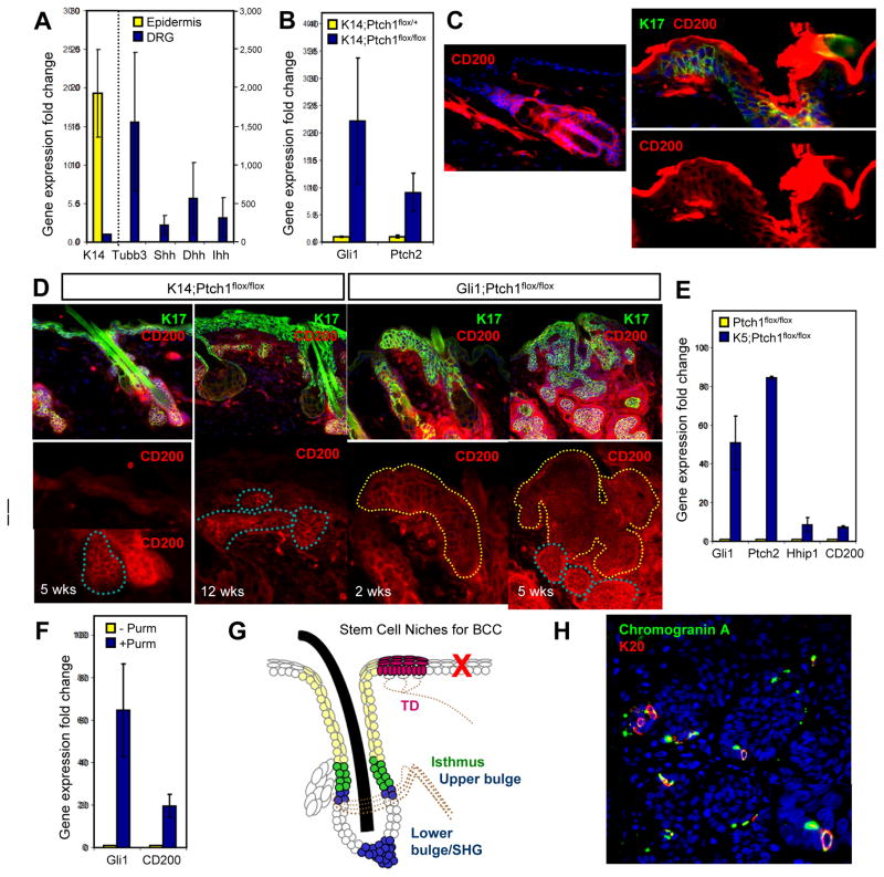
Basal cell carcinoma (BCC) is characterized by frequent loss of PTCH1, leading to constitutive activation of the Hedgehog pathway. Although the requirement for Hedgehog in BCC is well established, the identity of disease-initiating cells and the compartments in which they reside remain controversial. By using several inducible Cre drivers to delete Ptch1 in different cell compartments in mice, we show here that multiple hair follicle stem cell populations readily develop BCC-like tumors. In contrast, stem cells within the interfollicular epidermis do not efficiently form tumors. Notably, we observed that innervated Gli1-expressing progenitors within mechanosensory touch dome epithelia are highly tumorigenic. Sensory nerves activate Hedgehog signaling in normal touch domes, while denervation attenuates touch dome-derived tumors. Together, our studies identify varying tumor susceptibilities among different stem cell populations in the skin, highlight touch dome epithelia as "hot spots" for tumor formation, and implicate cutaneous nerves as mediators of tumorigenesis.


Figures

Figure 1
Figure 1. Multiple hair follicle stem cells readily form BCC-like tumors
A. Schematic showing areas of activity (blue) for the different inducible Cre recombinases used in this study. (*), TD epithelia. Yellow, sebaceous glands. B. Hematoxylin & eosin (H&E) staining showing that Gli1;Ptch1 mice, but not control animals, develop numerous hair follicle-associated tumors, 5 weeks after tamoxifen (TAM). C. Higher magnification views of hair follicle-associated tumors with peripheral palisading (dotted line). D. Hes1-CreERT2-mediated recombination of a floxed YFP reporter allele (green) in suprabasal cells of the epidermis, infundibulum and, less frequently, in the bulge, 3 days (left) or 50 days (right) post-TAM. E. Hes1;Ptch1 mice develop bulge-associated tumors, 7 weeks post-TAM. Right panel is a higher magnification view of (*). F. Lrig1;Ptch1 mice develop tumors associated with the isthmus and infundibulum, 5 weeks post-TAM. Scale bars, 50 μm. See also Figure S1.
Figure 2
Figure 2. IFE stem cells do not efficiently form tumors
A. IHC showing that K14-CreERT induces recombination of a YFP reporter allele (green) primarily in basal IFE cells, as marked by integrin β4 (red). B. K14;Ptch1 mice develop small ectopic IFE-associated buds, 5 weeks after TAM. By 12 weeks post-TAM, the IFE is hyperplastic but largely devoid of lesions. Tumors adjacent to the IFE (arrows) are typically connected to hair follicles, as shown in serial sections (Figure S2). C. Loss of p53 does not promote IFE tumor formation, 12 weeks post-TAM. Scale bars, 50 μm.
Figure 3
Figure 3. Hair follicle-derived tumors express similar markers regardless of cellular origin
IHC for (A) K14, (B) K17, (C) Sox9, (D) Lrig1, (E) K10, and (F) Involucrin (Inv). Wild-type telogen hair follicles were from 7.5 week old mice. Gli1;Ptch1 and Lrig1;Ptch1 tumors were collected 5 weeks post-TAM, while Hes1;Ptch1 and K14;Ptch1 samples were harvested 7 and 12 weeks post-TAM, respectively. Arrows indicate follicles where the bulge is visible. Scale bars, 50 μm.
Figure 4
Figure 4. TDs are hot spots for tumor formation
A. H&E staining of a TD-associated lesion (*) in Gli1;Ptch1 mice, 5 weeks post-TAM (left). Right, higher magnification view of (*). B. Top, whole mount LacZ staining of skin from Gli1;LacZ mice, 7 days (left) and 50 days (middle) post-TAM. Right, LacZ staining showing TD labeling. Bottom, IHC for YFP (green) and K17 (red) in a TD from a Gli1;YFP mouse, 35 days post-TAM. C. Resemblance of TD columnar basal cells with palisading periphery of a Gli1;Ptch1 tumor (dotted lines). D. Top left, IHC of a normal TD, identified by K17 expression (red) and underlying K8+ Merkel cells (green). Right, tumorigenic TDs from Gli1;Ptch1 mice, 2 and 5 weeks post-TAM. E. Quantitation of K17+ cells in the IFE and TD size in Gli1;Ptch1 mice or controls, 2 and 5 weeks post-TAM. F. Top, TD-derived K5+ tumor (red) retaining underlying Merkel cells (yellow) associated with nerves, as identified by Neurofilament (NF, green). Bottom, hair follicle-associated tumors (red) with nerves in the dermis (green), but no Merkel cells. G. K8+ Merkel cells (arrow) are associated with infrequent IFE-derived tumors, suggesting a TD origin. Inset, enlarged view. H. K14-CreERT displays infrequent recombination in K17+ TD epithelia (red), and frequent recombination in the rest of the IFE, as assessed by YFP reporter expression (green). I. Quantitation of Gli1-CreERT2 and K14-CreERT recombination rates in TD and non-TD IFE. NR, no recombination detected in non-TD IFE in Gli1;YFP mice. J. Reduced expression of K14 (red) in TDs overlying Merkel cells (green). Right panel is identical to the left, but with DAPI omitted. Data are represented as mean ± SEM. Scale bars, 50 μm.
Figure 5
Figure 5. Innervation is required for Hh signaling in TDs
A. IHC of denervated (“Den”) or sham-operated wild-type skin (“Sham”) showing TD epithelia (red) and Merkel cells (green), 5 weeks after surgery. Right, magnified views of TD areas (*). B. Quantitation of the average size of K17+ TDs, as well as the overall abundance of K17+ cells and Merkel cells in the IFE, 3 and 5 weeks after denervation. Matched sham and denervated data from the same mice are connected by lines. C. Denervation causes stable loss of nerve endings (red) from Merkel cells (green), 3 weeks after surgery. D. Whole-mount LacZ staining showing that denervation inhibits induction of LacZ reporter expression in Gli1;LacZ mice. Den, denervation; T, TAM; B, biopsy. All superimposed bar graphs depict mean values. Scale bars, 50 μm. See also Figure S3.
Figure 6
Figure 6. Denervation inhibits TD-derived tumors
A. Whole-mount LacZ staining of Gli1;LacZ skin, showing that denervation performed after TAM does not have an immediate effect on labeled Gli1-expressing TD cells (blue), 2 weeks after induction. B. IHC showing that denervation performed after TAM induction of Gli1;Ptch1 mice reduces the size and complexity of TD-derived tumors (red). K8+ Merkel cells are labeled green (arrowheads). C. Quantitation of TD-derived tumor cells (left panel) and Merkel cell abundance (right panel), 2 and 5 weeks post-TAM. Matched sham and denervated data from the same mice are connected by lines. D. H&E and quantitation showing that denervation does not affect the abundance of hair follicle-derived tumors. E. IHC and quantitation showing that denervation does not affect K14-CreERT-mediated recombination in the IFE. F. Quantitation showing that denervation inhibits tumor formation in TDs (left), but not the formation of ectopic IFE buds (right) in K14;Ptch1 mice. Den, denervation; T, TAM; B, biopsy. All superimposed bar graphs depict mean values. Data are represented as mean ± SEM. Scale bars, 50 μm. See also Figure S4.
Figure 7
Figure 7. A mechanosensory niche promotes tumorigenesis
A. qPCR showing that DRG (blue bars) highly express all 3 Hh ligands (Shh, Dhh, Ihh) relative to epidermis (yellow bars). K14 and Tubulin β3 (Tubb3) are specificity controls for skin and neurons, respectively. All values are expressed as fold induction relative to that of DRG (for K14, left y-axis) or epidermis (for all other genes, right y-axis). B. qPCR showing that K14;Ptch1 mice, relative to non-tumorigenic controls, upregulate Gli1 and Ptch2 in the skin, 5 weeks post-TAM. C. IHC showing CD200 expression (red) throughout the hair follicle (left) and TD (right, dotted line), but not in the IFE. Top right, TD marked by K17 (green) and CD200, or CD200 alone (bottom right). D. Left panels, IHC showing that CD200 (red) is highly expressed in hair follicle-derived tumors (blue dotted lines), but weakly expressed in ectopic IFE buds or hyperplastic IFE (white dotted lines) in K14;Ptch1 mice. Right panels, IHC showing that CD200 is highly expressed in TD-derived tumors (yellow dotted lines), both 2 and 5 weeks post-TAM, in Gli1;Ptch1 mice. K17 (green) is indiscriminately upregulated in hyperplastic IFE, ectopic IFE buds and tumors. All bottom panels are magnified views of the boxed areas. E. qPCR showing that keratinocytes harboring K5 promoter-driven constitutive Cre and homozygous Ptch1 flox alleles upregulate canonical Hh target genes as well as CD200. F. qPCR showing that the Smoothened agonist purmorphamine (purm) upregulates CD200 in wild-type keratinocytes. G. BCCs arise from multiple stem cell populations in the hair follicle and TD (filled regions). Tumors are also associated with the hair follicle infundibulum (beige), but do not arise efficiently from IFE stem cells (red ‘X’). Tumors associated with the middle bulge region were not observed in this study, possibly due to inefficient Cre-mediated recombination in this domain. Dotted lines, nerves. SHG, secondary hair germ. H. IHC showing Merkel cells (arrowheads) expressing Chromogranin A (green) and K20 (red) in a human BCC. Inset is a magnified view of the region identified by the arrow. Data are represented as mean ± SEM. Scale bars, 50 μm. See also Figures S5–S7.

Comment in

References

    1. Adolphe C, Nieuwenhuis E, Villani R, Li ZJ, Kaur P, Hui CC, Wainwright B. Patched1 and Patched2 redundancy plays a key role in regulating epidermal differentiation. J Invest Dermatol. 2014;134:1981–1990. - PubMed
    1. Ahn S, Joyner AL. Dynamic changes in the response of cells to positive hedgehog signaling during mouse limb patterning. Cell. 2004;118:505–516. - PubMed
    1. Aszterbaum M, Epstein J, Oro A, Douglas V, LeBoit PE, Scott MP, Epstein EH., Jr Ultraviolet and ionizing radiation enhance the growth of BCCs and trichoblastomas in patched heterozygous knockout mice. Nat Med. 1999;5:1285–1291. - PubMed
    1. Bastiaens MT, Hoefnagel JJ, Bruijn JA, Westendorp RG, Vermeer BJ, Bouwes Bavinck JN. Differences in age, site distribution, and sex between nodular and superficial basal cell carcinoma indicate different types of tumors. J Invest Dermatol. 1998;110:880–884. - PubMed
    1. Berton TR, Matsumoto T, Page A, Conti CJ, Deng CX, Jorcano JL, Johnson DG. Tumor formation in mice with conditional inactivation of Brca1 in epithelial tissues. Oncogene. 2003;22:5415–5426. - PubMed

Publication types

MeSH terms