

Beyond neurovascular coupling, role of astrocytes in the regulation of vascular tone
- PMID: 25843438
- PMCID: PMC4592693
- DOI: 10.1016/j.neuroscience.2015.03.064
Beyond neurovascular coupling, role of astrocytes in the regulation of vascular tone
Abstract
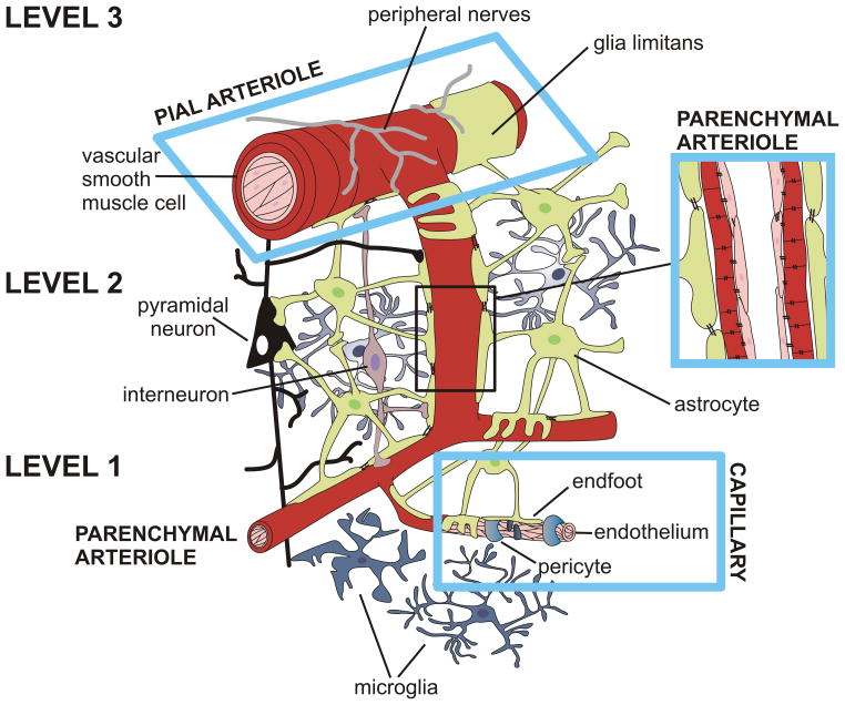
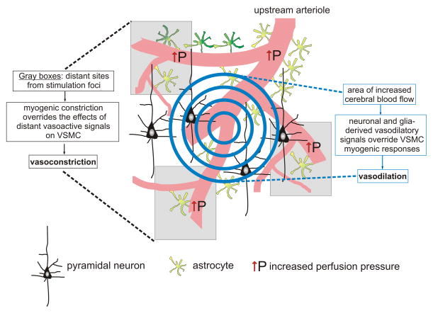
The brain possesses two intricate mechanisms that fulfill its continuous metabolic needs: cerebral autoregulation, which ensures constant cerebral blood flow over a wide range of arterial pressures and functional hyperemia, which ensures rapid delivery of oxygen and glucose to active neurons. Over the past decade, a number of important studies have identified astrocytes as key intermediaries in neurovascular coupling (NVC), the mechanism by which active neurons signal blood vessels to change their diameter. Activity-dependent increases in astrocytic Ca(2+) activity are thought to contribute to the release of vasoactive substances that facilitate arteriole vasodilation. A number of vasoactive signals have been identified and their role on vessel caliber assessed both in vitro and in vivo. In this review, we discuss mechanisms implicating astrocytes in NVC-mediated vascular responses, limitations encountered as a result of the challenges in maintaining all the constituents of the neurovascular unit intact and deliberate current controversial findings disputing a main role for astrocytes in NVC. Finally, we briefly discuss the potential role of pericytes and microglia in NVC-mediated processes.
Keywords: astrocyte; calcium; cerebral blood flow; myogenic tone; neurovascular coupling.
Copyright © 2015 IBRO. Published by Elsevier Ltd. All rights reserved.
Figures
Similar articles
-
Astrocyte regulation of cerebral vascular tone.Am J Physiol Heart Circ Physiol. 2013 Sep 1;305(5):H609-19. doi: 10.1152/ajpheart.00359.2013. Epub 2013 Jun 21. Am J Physiol Heart Circ Physiol. 2013. PMID: 23792684 Free PMC article. Review.
-
Astrocyte Ca2+ Signaling Drives Inversion of Neurovascular Coupling after Subarachnoid Hemorrhage.J Neurosci. 2015 Sep 30;35(39):13375-84. doi: 10.1523/JNEUROSCI.1551-15.2015. J Neurosci. 2015. PMID: 26424885 Free PMC article.
-
Tonic Local Brain Blood Flow Control by Astrocytes Independent of Phasic Neurovascular Coupling.J Neurosci. 2015 Sep 30;35(39):13463-74. doi: 10.1523/JNEUROSCI.1780-15.2015. J Neurosci. 2015. PMID: 26424891 Free PMC article.
-
Binaural blood flow control by astrocytes: listening to synapses and the vasculature.J Physiol. 2017 Mar 15;595(6):1885-1902. doi: 10.1113/JP270979. Epub 2016 Oct 14. J Physiol. 2017. PMID: 27619153 Free PMC article. Review.
-
Astrocyte contributions to flow/pressure-evoked parenchymal arteriole vasoconstriction.J Neurosci. 2015 May 27;35(21):8245-57. doi: 10.1523/JNEUROSCI.4486-14.2015. J Neurosci. 2015. PMID: 26019339 Free PMC article.
Cited by
-
Chronic cerebrovascular dysfunction after traumatic brain injury.J Neurosci Res. 2016 Jul;94(7):609-22. doi: 10.1002/jnr.23732. Epub 2016 Apr 27. J Neurosci Res. 2016. PMID: 27117494 Free PMC article. Review.
-
Impact of diabetes and ischemic stroke on the cerebrovasculature: A female perspective.Neurobiol Dis. 2022 Jun 1;167:105667. doi: 10.1016/j.nbd.2022.105667. Epub 2022 Feb 25. Neurobiol Dis. 2022. PMID: 35227927 Free PMC article. Review.
-
Research progress on the roles of neurovascular unit in stroke-induced immunosuppression.Zhejiang Da Xue Xue Bao Yi Xue Ban. 2023 Oct 25;52(5):662-672. doi: 10.3724/zdxbyxb-2023-0144. Zhejiang Da Xue Xue Bao Yi Xue Ban. 2023. PMID: 37899404 Free PMC article. Review. Chinese, English.
-
Fractalkine-induced microglial vasoregulation occurs within the retina and is altered early in diabetic retinopathy.Proc Natl Acad Sci U S A. 2021 Dec 21;118(51):e2112561118. doi: 10.1073/pnas.2112561118. Proc Natl Acad Sci U S A. 2021. PMID: 34903661 Free PMC article.
-
Astrocyte dysfunction and neurovascular impairment in neurological disorders: Correlation or causation?Neurochem Int. 2019 Sep;128:70-84. doi: 10.1016/j.neuint.2019.04.005. Epub 2019 Apr 13. Neurochem Int. 2019. PMID: 30986503 Free PMC article. Review.
References
-
- Abudara V, Roux L, Dallerac G, Matias I, Dulong J, Mothet JP, Rouach N, Giaume C. Activated microglia impairs neuroglial interaction by opening Cx43 hemichannels in hippocampal astrocytes. Glia 2015 - PubMed
-
- Alkayed NJ, Narayanan J, Gebremedhin D, Medhora M, Roman RJ, Harder DR. Molecular characterization of an arachidonic acid epoxygenase in rat brain astrocytes. Stroke. 1996;27:971–979. - PubMed
-
- Alonso-Galicia M, Hudetz AG, Shen H, Harder DR, Roman RJ. Contribution of 20-HETE to vasodilator actions of nitric oxide in the cerebral microcirculation. Stroke. 1999;30:2727–2734. discussion 2734. - PubMed
-
- Anderson CM, Bergher JP, Swanson RA. ATP-induced ATP release from astrocytes. J Neurochem. 2004;88:246–256. - PubMed
Publication types
MeSH terms
Grants and funding
LinkOut - more resources
Full Text Sources
Other Literature Sources
Miscellaneous
