. 2015;9(4):3926-34.
doi: 10.1021/nn507241v. Epub 2015 Apr 8.

In Vivo Tumor Vasculature Targeting of CuS@MSN Based Theranostic Nanomedicine

Affiliations

In Vivo Tumor Vasculature Targeting of CuS@MSN Based Theranostic Nanomedicine

Feng Chen et al. ACS Nano. 2015.

Abstract

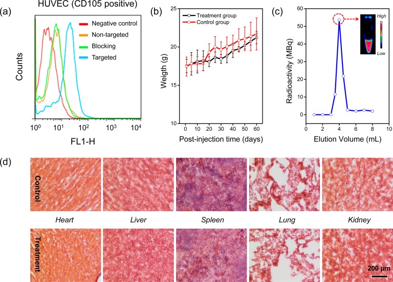
Actively targeted theranostic nanomedicine may be the key for future personalized cancer management. Although numerous types of theranostic nanoparticles have been developed in the past decade for cancer treatment, challenges still exist in the engineering of biocompatible theranostic nanoparticles with highly specific in vivo tumor targeting capabilities. Here, we report the design, synthesis, surface engineering, and in vivo active vasculature targeting of a new category of theranostic nanoparticle for future cancer management. Water-soluble photothermally sensitive copper sulfide nanoparticles were encapsulated in biocompatible mesoporous silica shells, followed by multistep surface engineering to form the final theranostic nanoparticles. Systematic in vitro targeting, an in vivo long-term toxicity study, photothermal ablation evaluation, in vivo vasculature targeted imaging, biodistribution and histology studies were performed to fully explore the potential of as-developed new theranostic nanoparticles.

Keywords: CuS; mesoporous silica nanoparticle; photothermal ablation; theranostic nanomedicine; vasculature targeting.


Figures

Figure 1
Figure 1
Synthesis and characterization of CuS@MSN. (a) A schematic illustration showing 3 major steps for the synthesis of 64Cu-CuS@MSN-TRC105 theranostic nanoparticle. Step 1: encapsulating CuS-CTAC inside mesoporous silica shell, resulting in CuS@MSN nanoparticle. Step 2: surface engineering of CuS@MSN to form 64Cu-CuS@MSN-TRC105 nanoconjugates (see Figure S1 for more details). (b) A TEM image of CuS-CTAC. Inset shows the digital photo of CuS-CTAC in water. (c) A TEM image of CuS@MSN. Inset shows the digital photo of CuS@MSN in water. (d) Nitrogen adsorption–desorption isotherms and the corresponding pore size distribution of CuS@MSN (inset, see Figure S4 for enlarged figure). (e) UV–vis absorption spectra of CuS-CTAC (black line) and CuS@MSN (red line). Inset shows the photothermal images of water, CuS-CTAC and CuS@MSN (from left to right) (see Figure S5 for enlarged figure). Note, both CuS-CTAC and CuS@MSN have the same amount of CuS nanoparticles (i.e., 0.25 mM of Cu based on ICP-AES measurement). (f) Quantitative temperature change of CuS@MSN aqueous solution as a function of 980 nm laser exposure time (laser power density: 4.0 W/cm2). Concentration of CuS@MSN was based on total Cu amount measured by using ICP-AES. Inset shows the increased temperature for samples with varied CuS@MSN concentrations (see Figure S6 for enlarged figure).
Figure 2
Figure 2
In vivo photothermal therapeutic evaluation. Photothermal images of mice after laser treatment. (a) (CuS@MSN+980 nm laser) group, (b) (980 nm laser only) group, and (c) (CuS@MSN only) group. Digital photos of mice with 4T1 tumors on Day 30 after treatment. (d) (CuS@MSN+980 nm laser) group, (e) (980 nm laser only) group, and (f) (CuS@MSN only) group. (g) A digital photo of mouse from (CuS@MSN+980 nm laser) group on Day 67 after treatment. (h) Changes of tumor size of mice from 3 different groups after treatment (n = 5). Laser dose: 4 W/cm2, 15 min. CuS@MSN dose: 33 mg/kg. Tumors were marked with red arrows. The differences between the treatment group and two control groups were statistically significant (*P < 0.05 on Day 4, **P < 0.01 at later time points).
Figure 3
Figure 3
In vitro CD105 targeting, long-term toxicity and radiolabeling studies. (a) Flow cytometry analysis of CuS@MSN nanoconjugates in HUVEC (CD105 positive) cell lines (incubation time was set at 30 min). (b) Two-month growth chart of mice from treatment (dose: 90 mg/kg of PEGylated CuS@MSN in PBS) and control groups (PBS only). (c) A size-exclusion column chromatography elution profile during the purification of 64Cu-CuS@MSN-TRC105. The unreacted 64Cu elutes after 6 mL. Inset shows the PET imaging of fraction 3.5–4.0 mL. (d) H&E-stained tissue sections from mice injected with PEGylated CuS@MSN (treatment group) and PBS only (control group) after 60 days. Tissues were harvested from heart, liver, spleen, lung and kidney.
Figure 4
Figure 4
In vivo CD105 targeted PET imaging, tumor uptake comparison and histology studies. In vivo serial coronal PET images of 64Cu-CuS@MSN-TRC105 nanoconjugates (a, targeted group), 64Cu-CuS@MSN (b, nontargeted group) and 64Cu-CuS@MSN-TRC105 with a large dose of free TRC105 (c, blocking group) in 4T1 murine breast tumor-bearing mice at different time points postinjection. (d) Tumor uptake comparison among 3 different groups. The difference between 4T1 tumor uptake in targeted group and two control groups were statistically significant (**P < 0.01). (e) Ex vivo histology analysis of the tumor tissue slices with CD31 (red, with antimouse CD31 primary antibody) and CD105 (green, using the TRC105 from 64Cu-CuS@MSN-TRC105 as the primary antibody). Muscle slices were also provided. Tumors were marked with yellow arrows.

References

    1. Jokerst J. V.; Gambhir S. S. Molecular Imaging with Theranostic Nanoparticles. Acc. Chem. Res. 2011, 44, 1050–1060. - PMC - PubMed
    1. Blackwell K. L.; Burstein H. J.; Storniolo A. M.; Rugo H. S.; Sledge G.; Aktan G.; Ellis C.; Florance A.; Vukelja S.; Bischoff J.; et al. Overall Survival Benefit with Lapatinib in Combination with Trastuzumab for Patients with Human Epidermal Growth Factor Receptor 2-Positive Metastatic Breast Cancer: Final Results from the EGF104900 Study. J. Clin. Oncol. 2012, 30, 2585–2592. - PubMed
    1. Benezra M.; Penate-Medina O.; Zanzonico P. B.; Schaer D.; Ow H.; Burns A.; DeStanchina E.; Longo V.; Herz E.; Iyer S.; et al. Multimodal Silica Nanoparticles Are Effective Cancer-Targeted Probes in a Model of Human Melanoma. J. Clin. Invest. 2011, 121, 2768–2780. - PMC - PubMed
    1. Chen F.; Cai W. Tumor Vasculature Targeting: A Generally Applicable Approach for Functionalized Nanomaterials. Small 2014, 10, 1887–1893. - PMC - PubMed
    1. Xie J.; Lee S.; Chen X. Nanoparticle-Based Theranostic Agents. Adv. Drug Delivery Rev. 2010, 62, 1064–1079. - PMC - PubMed

Publication types

MeSH terms