

Contribution of protease-activated receptor 1 in status epilepticus-induced epileptogenesis
- PMID: 25843668
- PMCID: PMC4682556
- DOI: 10.1016/j.nbd.2015.03.026
Contribution of protease-activated receptor 1 in status epilepticus-induced epileptogenesis
Abstract
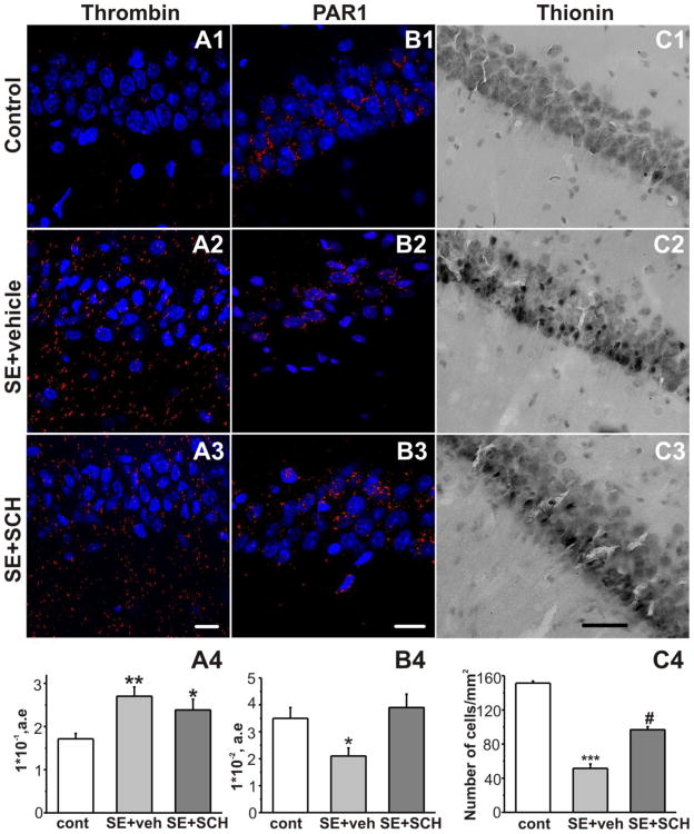
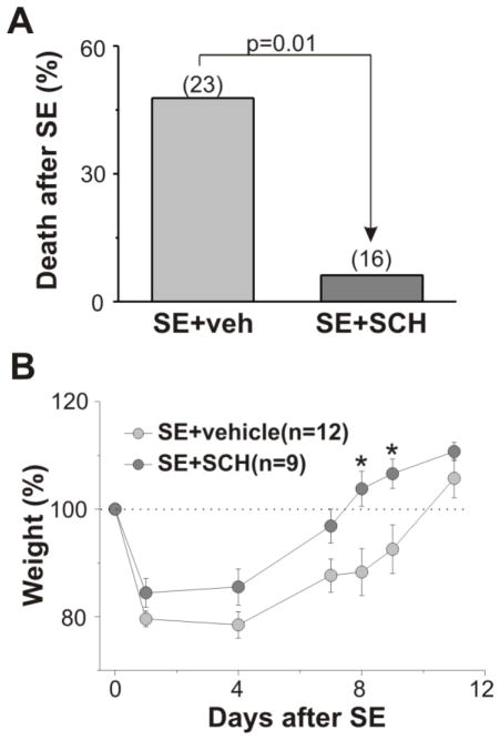
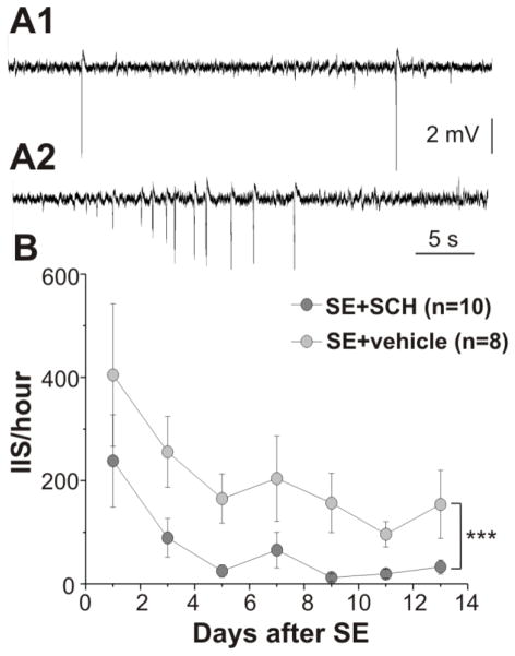
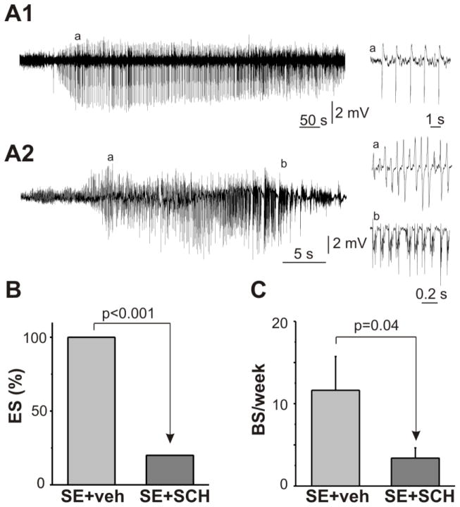
Clinical observations and studies on different animal models of acquired epilepsy consistently demonstrate that blood-brain barrier (BBB) leakage can be an important risk factor for developing recurrent seizures. However, the involved signaling pathways remain largely unclear. Given the important role of thrombin and its major receptor in the brain, protease-activated receptor 1 (PAR1), in the pathophysiology of neurological injury, we hypothesized that PAR1 may contribute to status epilepticus (SE)-induced epileptogenesis and that its inhibition shortly after SE will have neuroprotective and antiepileptogenic effects. Adult rats subjected to lithium-pilocarpine SE were administrated with SCH79797 (a PAR1 selective antagonist) after SE termination. Thrombin and PAR1 levels and neuronal cell survival were evaluated 48h following SE. The effect of PAR1 inhibition on animal survival, interictal spikes (IIS) and electrographic seizures during the first two weeks after SE and behavioral seizures during the chronic period was evaluated. SE resulted in a high mortality rate and incidence of IIS and seizures in the surviving animals. There was a marked increase in thrombin, decrease in PAR1 immunoreactivity and hippocampal cell loss in the SE-treated rats. Inhibition of PAR1 following SE resulted in a decrease in mortality and morbidity, increase in neuronal cell survival in the hippocampus and suppression of IIS, electrographic and behavioral seizures following SE. These data suggest that the PAR1 signaling pathway contributes to epileptogenesis following SE. Because breakdown of the BBB occurs frequently in brain injuries, PAR1 inhibition may have beneficial effects in a variety of acquired injuries leading to epilepsy.
Keywords: Epileptogenesis; Hippocampus; Pilocarpine; Protease-activated receptor; Status epilepticus; Thrombin.
Copyright © 2015 Elsevier Inc. All rights reserved.
Figures
Similar articles
-
Systemic thrombin inhibition ameliorates seizures in a mouse model of pilocarpine-induced status epilepticus.J Mol Med (Berl). 2019 Nov;97(11):1567-1574. doi: 10.1007/s00109-019-01837-2. Epub 2019 Oct 31. J Mol Med (Berl). 2019. PMID: 31667526
-
Inhibition of protease-activated receptor 1 ameliorates behavioral deficits and restores hippocampal synaptic plasticity in a rat model of status epilepticus.Neurosci Lett. 2019 Jan 23;692:64-68. doi: 10.1016/j.neulet.2018.10.058. Epub 2018 Nov 1. Neurosci Lett. 2019. PMID: 30391321
-
Effects of protease-activated receptor 1 inhibition on anxiety and fear following status epilepticus.Epilepsy Behav. 2017 Feb;67:66-69. doi: 10.1016/j.yebeh.2016.11.003. Epub 2017 Jan 13. Epilepsy Behav. 2017. PMID: 28088683
-
Neurocoagulation from a Mechanistic Point of View in the Central Nervous System.Semin Thromb Hemost. 2022 Apr;48(3):277-287. doi: 10.1055/s-0041-1741569. Epub 2022 Jan 20. Semin Thromb Hemost. 2022. PMID: 35052009 Review.
-
TRPC Channels and Epilepsy.Adv Exp Med Biol. 2017;976:123-135. doi: 10.1007/978-94-024-1088-4_11. Adv Exp Med Biol. 2017. PMID: 28508318 Review.
Cited by
-
Leukocyte differential gene expression prognostic value for high versus low seizure frequency in temporal lobe epilepsy.BMC Neurol. 2024 Jan 2;24(1):16. doi: 10.1186/s12883-023-03459-1. BMC Neurol. 2024. PMID: 38166692 Free PMC article.
-
Targeting prostaglandin receptor EP2 for adjunctive treatment of status epilepticus.Pharmacol Ther. 2020 May;209:107504. doi: 10.1016/j.pharmthera.2020.107504. Epub 2020 Feb 21. Pharmacol Ther. 2020. PMID: 32088247 Free PMC article. Review.
-
A Linear Temporal Increase in Thrombin Activity and Loss of Its Receptor in Mouse Brain following Ischemic Stroke.Front Neurol. 2017 Apr 10;8:138. doi: 10.3389/fneur.2017.00138. eCollection 2017. Front Neurol. 2017. PMID: 28443061 Free PMC article.
-
Systemic thrombin inhibition ameliorates seizures in a mouse model of pilocarpine-induced status epilepticus.J Mol Med (Berl). 2019 Nov;97(11):1567-1574. doi: 10.1007/s00109-019-01837-2. Epub 2019 Oct 31. J Mol Med (Berl). 2019. PMID: 31667526
-
Status Epilepticus Induced Spontaneous Dentate Gyrus Spikes: In Vivo Current Source Density Analysis.PLoS One. 2015 Jul 6;10(7):e0132630. doi: 10.1371/journal.pone.0132630. eCollection 2015. PLoS One. 2015. PMID: 26148195 Free PMC article.
References
-
- Baraban JM, Fiore RS, Sanghera JS, Paddon HB, Pelech SL. Identification of p42 mitogen-activated protein kinase as a tyrosine kinase substrate activated by maximal electroconvulsive shock in hippocampus. J Neurochem. 1993;60:330–6. - PubMed
-
- Berkeley JL, Decker MJ, Levey AI. The role of muscarinic acetylcholine receptor-mediated activation of extracellular signal-regulated kinase 1/2 in pilocarpine-induced seizures. J Neurochem. 2002;82:192–201. - PubMed
-
- Bourgognon JM, Schiavon E, Salah-Uddin H, Skrzypiec aE, Attwood BK, Shah RS, Patel SG, Mucha M, John Challiss Ra, Forsythe ID, Pawlak R. Regulation of neuronal plasticity and fear by a dynamic change in PAR1-G protein coupling in the amygdala. Mol Psychiatry. 2013;18:1136–45. doi: 10.1038/mp.2012.133. - DOI - PMC - PubMed
Publication types
MeSH terms
Substances
Grants and funding
LinkOut - more resources
Full Text Sources
Other Literature Sources
