. 2015 Apr 14;11(2):249-60.
doi: 10.1016/j.celrep.2015.03.016. Epub 2015 Apr 2.

Casein kinase 1δ is an APC/C(Cdh1) substrate that regulates cerebellar granule cell neurogenesis

Affiliations

Casein kinase 1δ is an APC/C(Cdh1) substrate that regulates cerebellar granule cell neurogenesis

Clara Penas et al. Cell Rep. .

Abstract

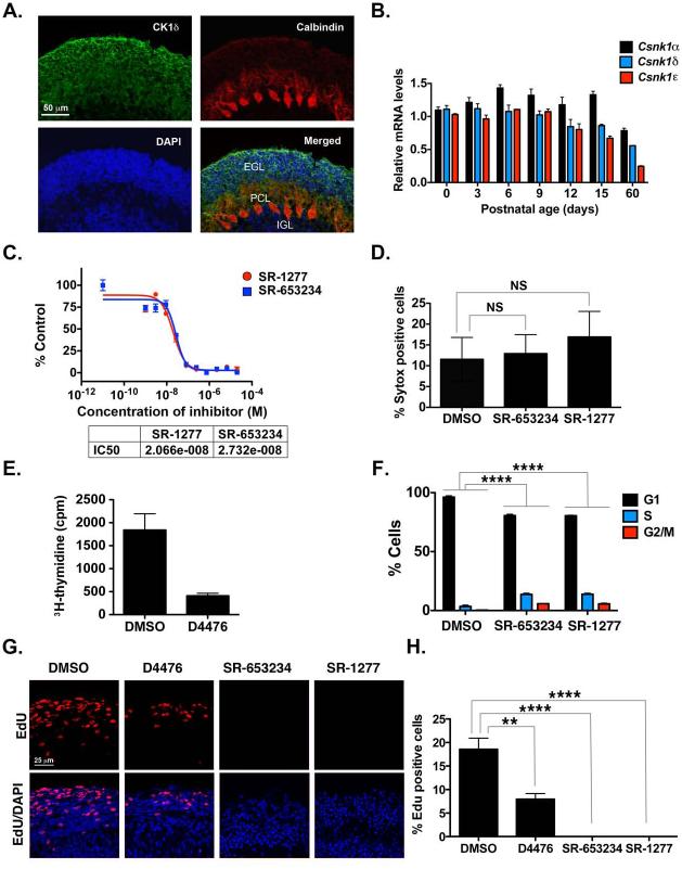
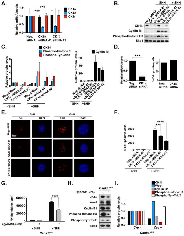
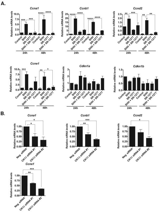
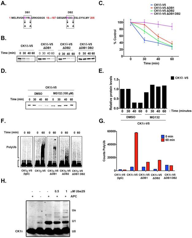
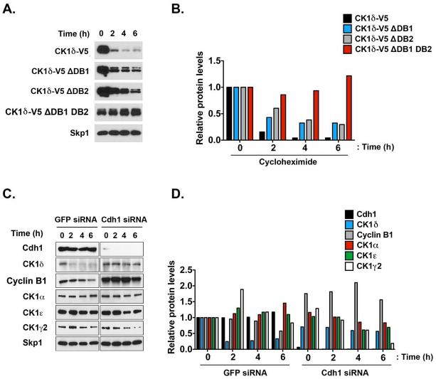
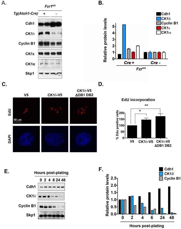
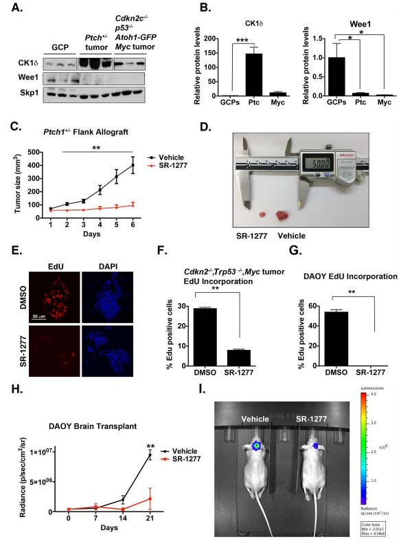
Although casein kinase 1δ (CK1δ) is at the center of multiple signaling pathways, its role in the expansion of CNS progenitor cells is unknown. Using mouse cerebellar granule cell progenitors (GCPs) as a model for brain neurogenesis, we demonstrate that the loss of CK1δ or treatment of GCPs with a highly selective small molecule inhibits GCP expansion. In contrast, CK1δ overexpression increases GCP proliferation. Thus, CK1δ appears to regulate GCP neurogenesis. CK1δ is targeted for proteolysis via the anaphase-promoting complex/cyclosome (APC/C(Cdh1)) ubiquitin ligase, and conditional deletion of the APC/C(Cdh1) activator Cdh1 in cerebellar GCPs results in higher levels of CK1δ. APC/C(Cdh1) also downregulates CK1δ during cell-cycle exit. Therefore, we conclude that APC/C(Cdh1) controls CK1δ levels to balance proliferation and cell-cycle exit in the developing CNS. Similar studies in medulloblastoma cells showed that CK1δ holds promise as a therapeutic target.


Figures

Figure 1
Figure 1. CK1δ Expression in Postnatal GCPs and Control of GCP Proliferation in vitro and ex vivo
(A) Cerebellar sections from P8 pups were stained with antibodies against CK1δ (green) or calbindin (red) and DAPI (blue). (B) CK1α , CK1δ, and CK1ε mRNA was amplified by qRT-PCR, and fold-change in gene expression in postnatal mouse cerebellum was determined by normalizing to GAPDH values relative to control. (C) GCPs were incubated for 24 h with increasing concentrations of SR-653234 or SR-1277, and the amount of proliferation was determined by 3H-thymidine incorporation. Results were plotted relative to that seen in the DMSO control. (D) GCPs were treated with 100 nM SR-653234 or SR-1277 for 24 h, and then Sytox and Hoechst staining was performed (NS, not significant, as determined by one-way ANOVA and Dunnett multiple comparisions test). (E) D4476 (20 μM) reduces GCP proliferation, and (F) SR-653234 (100 nM) and SR-1277 (100 nM) increase the percentage of GCPs in the S or G2/M phase. GCPs were treated for 24 h with the indicated compounds or DMSO, and the proportion of cells in each cell cycle phase was determined by PI-FACS. (G) Organotypic cerebellar slices were treated with SR-1277 (100 nM), SR-653234 (100 nM), D4476 (20 μM), or DMSO for 1 h, after which EdU was added to the media for 20 h. Slices were stained with EdU (red) and the nuclear marker DAPI (blue). (H) Quantification of (G). Results are shown as the average values of three independent experiments and are represented as the mean and standard error of the mean (± SEM) (*p <0.05, **p <0.001, ***p <0.001, ****p <0.0001).
Figure 2
Figure 2. CK1δ Knockdown Reduces GCP Proliferation in vitro and ex vivo
(A) GCP cells electroporated with two different CK1δ siRNAs were analyzed by qRT-PCR. CK1δ was significantly knocked down, but CK1α and CK1ε levels were not altered. The mRNA was amplified by qRT-PCR, and the fold-change in gene expression was determined by normalizing to GAPDH values relative to controls. (B) After electroporation of GPCs with or without SHH (75 ng/mL), the levels of CK1δ, cyclin B1, and phospho–histone H3 were analyzed by immunoblotting with antibodies against the proteins. Skp1 served as a loading control. CK1δ was knocked down by both siRNAs. (C) Quantification of the amount of CK1δ, phospho-Histone H3, phospho-Tyr-Cdk1 (a measure of Wee1 inhibition of Cdk1), and cyclin B1, relative to the loading control Skp1, after CK1δ siRNA electroporation in GCPs from (B). (D) GCPs electroporated with CK1ε siRNA were analyzed by qRT-PCR. CK1ε was significantly knocked down, but EdU uptake was not reduced in the presence of SHH. Levels are expressed as the percentage of EdU uptake in cells treated with the negative siRNA. (E) CK1δ siRNA electroporation reduces EdU uptake of GCPs in the presence of SHH. Proliferative GCP aggregates were stained with EdU (red) and the nuclear marker DAPI (blue). (F) Quantification of the EdU incorporation in (E). Levels are expressed as a percentage compared to that in cells treated with the negative siRNA without SHH. (G) GCPs isolated from Csnk1d-deleted mice proliferate less in the presence or absence of SHH. Purified GCPs were treated for 24 h with the compounds, and then 3H-thymidine was added to the media for an additional 24 h. Plots representing the amounts of 3H-thymidine incorporated by GCPs from CK1δ-deleted or control mice are shown. (H) GCPs purified from conditional Csnk1d-deleted mice have lower levels of CK1δ, cyclin B1, and phospho-Histone H3, indicating decreased proliferation. Representative immunoblots of CK1δ, Wee1, cyclin B1, phospho-Histone H3, and phospho-Tyr-cdc2 relative to Skp1 are shown. (I) Quantification of the amount of CK1δ , Wee1, phospho-Histone H3, phospho-Tyr-Cdc2, and cyclin B1 protein, relative to the loading control Skp1, in GCPs isolated from Tg(Atoh1-Cre)+;Csnk1dfl/fl or Tg (Atoh1-Cre)-;Csnk1dfl/fl mice from (H). Results are shown as the averages of three independent experiments and are represented as the mean ± SEM (***p <0.001, ****p <0.0001).
Figure 3
Figure 3. Inhibition or Knockdown of CK1δ Reduces the mRNA Levels of Cell Cycle Components
(A) SR-1277 (100 nM) decreases the expression of SHH-induced levels of Ccna1, Ccnb1, Ccnd2, Ccne1, Cdkn1a, and Cdkn1b mRNA in GCPs. GCPs were treated with SHH (75 ng/mL) and/or SR-1277 for 24 or 48 h. The mRNA was amplified by qRT-PCR, and fold-change in gene expression was determined by normalizing to GAPDH values relative to control samples. (B) CK1δ knockdown reduces the expression of Ccna1, Ccnb1, Ccnd2, and Ccne1 mRNA levels in the presence of SHH. GCPs were electroporated with two different siRNAs against CK1δ, and the mRNA levels were analyzed after 72 h in vitro. Results shown are the averages of three independent experiments and are represented as the mean ± SEM (*p <0.05, **p <0.001, ***p <0.001, ****p <0.0001).
Figure 4
Figure 4. APC/CCdh1 induces CK1δ Ubiquitination and Degradation in vitro
(A) Two destruction boxes (D-boxes) in human CK1δ, DB1 and DB2, were mutated by substituting alanine (A) for the corresponding arginine (R) and leucine (L) residues. (B-C) Both D-boxes in CK1δ are required for proteolysis. In vitro degradation assay showing 35S-labeled wild-type CK1δ-V5, DB1 mutant (CK1δ-V5 ΔDB1), DB2 mutant (CK1δ-V5 ΔDB2), and DB1 DB2 double-mutant (CK1δ-V5 ΔDB1 DB2) after incubation in extracts prepared from HeLa cells in G1. Samples collected at the indicated time points were analyzed by autoradiography. (C) The quantification of (B); protein levels were measured in three separate experiments using Quantity One image analysis software (Bio-Rad). An unpaired t test was performed, and a p-value of 0.01 was obtained. (D) Autoradiogram showing in vitro degradation of 35S-labeled wild-type CK1δ-V5 in HeLa cell extracts at G1, in the presence or absence of the proteasome 26S inhibitor MG132 (100 μM). (E) Quantification of CK1δ-V5 from (D). (F-G) Both D-boxes in CK1δ are required for efficient ubiquitination. Autoradiogram of 35S-labeled wild-type CK1δ-V5, ΔDB1, ΔDB2, and ΔDB1 DB2 mutants after in vitro ubiquitination by anti-Cdc27 immunoprecipitates from HeLa cell extracts at G1. (G) The extent of polyubiquitination was quantified for the entire lane above the inputs by using Quantity One image analysis software. From three separate experiments, an unpaired t test was performed, and a p-value of 0.005 was obtained. Results shown are the averages of three independent experiments and are represented as the mean ± SEM. (H) Purified CK1δ and immunoprecipitated APC/C were incubated together in vitro, and the extent of ubiquitination was determined after SDS-PAGE and anti-CK1δ autoradiography.
Figure 5
Figure 5. CK1δ is Degraded by APC/CCdh1 in a D-box–dependent Manner
(A) CK1δ D-box mutations reduce the turnover of the protein in HeLa cells. HeLa cells transfected with the wild-type CK1δ-V5 or D-box mutants were treated with cycloheximide (100 μg/mL). Samples were then collected at the indicated time points and analyzed by immunoblotting. (B) Quantification of (A). (C) Cdh1 is required for CK1δ degradation. HeLa cells were transfected with the indicated siRNA, treated with cycloheximide for the indicated times, retrieved at the time points shown, and analyzed by immunoblotting. (D) Quantification of (C).
Figure 6
Figure 6. Conditional Deletion of Fzr1 in the Cerebellum Increases CK1δ Levels
(A) Immunoblot analysis showing that the levels of cyclin B1 and CK1δ, but not CK1ε, are higher in GCPs purified from Fzr1-knockout mice than in those from control mice. Protein extracts were made directly after GCP purification. GCPs were not maintained in culture. (B) Quantification of (A). (C) Overexpression of CK1δ-V5 in purified GCPs increases cell proliferation, as indicated by the amount of EdU-positive cells (red) in the presence of SHH (75 ng/mL). EdU incorporation into cells electroporated with the CK1δ-V5 or CK1δ-V5 ΔDB1 DB2 construct was normalized to that of cells electroporated with the empty control vector (V5). (D) Quantification of (C). (E) CK1δ levels decrease during GCP cell cycle exit. Representative Western blotting of CK1δ, Cdh1, cyclin B1, and the loading control Skp1. (F) Quantification of (E). Results shown are averages of three independent experiments and are represented as the mean ± SEM (*p <0.05, **p <0.001).
Figure 7
Figure 7. Murine Medulloblastoma Cells Express Elevated Levels of CK1δ, the Inhibition of which Reduces Tumor Growth in vivo
(A) CK1δ protein is overexpressed in Ptch1+/−, Cdkn2−/−, Trp53−/−, and c-Myc tumors, whereas Wee1 is downregulated. Skp1 was used as a loading control. (B) Quantification of (A). (C-D) SR-1277 decreases proliferation of Ptch1+/− allograft tumors. Ptch1+/− tumor cells were injected subcutaneously into mice. Once the tumor reached a volume of 50 to 90 mm3, treatment with vehicle or SR-1277 (20 mg/kg, twice daily) was initiated. Tumor size was quantified in four samples for each time point, and the averages are shown. (D) An image showing representative SR-1277– treated (left) and vehicle-treated (right) tumors. (E) Proliferation of Cdkn2−/−, Trp53−/−, and c-Myc tumor cells is reduced in the presence of SR-1277. (F) Quantification of EdU incorporation into Cdkn2−/−, Trp53−/−, or c-Myc tumor cells after DMSO or SR-1277 treatment. (G) EdU-incorporation assay shows that proliferation of DAOY cells is reduced in the presence of SR-1277 (500 nM). (H-I) SR-1277 also reduces the intracranial growth of DAOY cells. (H) Twelve days after mice were transplanted with DAOY tumor cells, D-luciferin was administered intraperitoneally, and bioluminescence was measured. (I) Fluorescence imaging of representative mice in which DAOY cells were implanted intracraniallly and then treated with SR-1277 (20 mg/kg, twice daily) or vehicle for 21 days. Bioluminescence was quantified from the encircled regions that enclose the entire tumor. Results shown are the means ± SEM of three independent experiments (*p <0.05, **p <0.001, ***p <0.001).

References

    1. Barford D. Structure, function and mechanism of the anaphase promoting complex (APC/C) Q. Rev. Biophys. 2011;44:153–190. - PubMed
    1. Behrend L, Stoter M, Kurth M, Rutter G, Heukeshoven J, Deppert W, Knippschild U. Interaction of casein kinase 1 delta (CK1delta) with post-Golgi structures, microtubules and the spindle apparatus. Eur. J. C.ell Biol. 2000;79:240–251. - PubMed
    1. Ben-Arie N, Bellen HJ, Armstrong DL, McCall AE, Gordadze PR, Guo Q, Matzuk MM, Zoghbi HY. Math1 is essential for genesis of cerebellar granule neurons. Nature. 1997;390:169–172. - PubMed
    1. Beyaert R, Vanhaesebroeck B, Declercq W, Van Lint J, Vandenabele P, Agostinis P, Vandenheede JR, Fiers W. Casein kinase-1 phosphorylates the p75 tumor necrosis factor receptor and negatively regulates tumor necrosis factor signaling for apoptosis. J. Biol. Chem. 1995;270:23293–23299. - PubMed
    1. Bibian M, Rahaim RJ, Choi JY, Noguchi Y, Schurer S, Chen W, Nakanishi S, Licht K, Rosenberg LH, Li L, et al. Development of highly selective casein kinase 1delta/1epsilon (CK1delta/epsilon) inhibitors with potent antiproliferative properties. Bioorg. Med. Chem. Lett. 2013;23:4374–4380. - PMC - PubMed

Publication types

MeSH terms

LinkOut - more resources