Review
. 2015 Jul;27(7):1380-91.
doi: 10.1016/j.cellsig.2015.03.018. Epub 2015 Apr 2.

microRNA regulation of Wnt signaling pathways in development and disease

Affiliations
Review

microRNA regulation of Wnt signaling pathways in development and disease

Jia L Song et al. Cell Signal. 2015 Jul.

Abstract

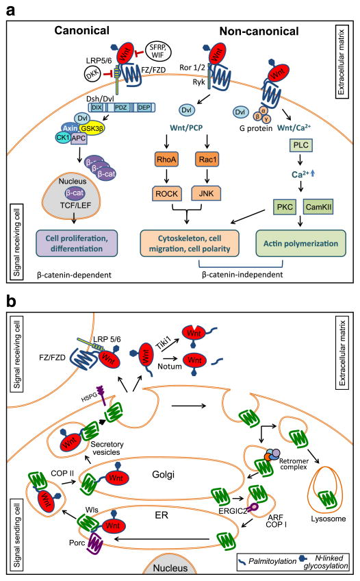
Wnt signaling pathways and microRNAs (miRNAs) are critical regulators of development. Aberrant Wnt signaling pathways and miRNA levels lead to developmental defects and diverse human pathologies including but not limited to cancer. Wnt signaling pathways regulate a plethora of cellular processes during embryonic development and maintain homeostasis of adult tissues. A majority of Wnt signaling components are regulated by miRNAs which are small noncoding RNAs that are expressed in both animals and plants. In animal cells, miRNAs fine tune gene expression by pairing primarily to the 3'untranslated region of protein coding mRNAs to repress target mRNA translation and/or induce target degradation. miRNA-mediated regulation of signaling transduction pathways is important in modulating dose-sensitive response of cells to signaling molecules. This review discusses components of the Wnt signaling pathways that are regulated by miRNAs in the context of development and diseases. A fundamental understanding of miRNA functions in Wnt signaling transduction pathways may yield new insight into crosstalks of regulatory mechanisms essential for development and disease pathophysiology leading to novel therapeutics.

Keywords: Cancer; Non-canonical Wnt signaling; Post-transcriptional regulation; Regulatory network; β-catenin.


Conflict of interest statement

Conflict of interest statement

The authors declare no conflict of interests.

Figures

Fig. 1
Fig. 1
Wnt signaling pathways. a) In canonical Wnt pathway, β-catenin is normally degraded by the destruction complex composed of Axin, APC, CK1, and GSK3β. Upon Wnt ligand binding to the frizzled (FZ/FDZ) and LRP5/6 co-receptor, disheveled (Dsh/Dvl) disassembles the β-catenin destruction complex, resulting in an accumulation of β-catenin that can enter the nucleus. In conjunction with TCF/LEF, β-catenin transcriptionally activates target genes. In non-canonical Wnt/Planar cell polarity pathway (Wnt/PCP), its activation is mediated by Wnt ligand binding to the FZ/FDZ and co-receptors Ror1, Ror2, or Ryk. Wnt/PCP is mediated by small GTPases RhoA and Rac1 to activate JNK and ROCK pathways, respectively, resulting in actin remodeling events. In Wnt/Ca2+ pathway, the activation of FZ/FDZ is mediated by disheveled and heterotrimetic G-proteins. Calcium release activates PKC, calcineurin, and CamKII that activates cell migration. b) Wnt proteins are N-glycosylated and lipid modified by the oligosacchary transferase complex and acyltransferase Porcupine within the endoplasmic reticulum (ER), respectively. Wnts are then engaged by Wntless (Wls) for secretory pathway transit and extracellular secretion. Wnt-Wls complex is transferred from ER to Golgi in COP II regulated vesicles, and from the Golgi to extracellular matrix in secretory vesicles [351,352]. Secretion mechanisms of Wnts are not completely understood; however, it has been reported that Heparan Sulfate Proteoglycans (HSPGs) are involved in Wnt stabilization in extracellular matrix [353]. After secretion, the Wnt ligand may be enzymatically inactivated by Tiki1 or Notum. Tiki1 is a protease that cleaves the amino terminal residues of Wnt [141], and Notum removes an essential palmitoleate moiety from Wnt [143]. Functional Wnts are then targeted to their receptors (FZ/FZD) via one of the proposed models mentioned in the text. Following extracellular release of Wnts, Wls is routed to multicellular vesicles for lysosomal degradation or shuttled back to the Golgi by retromer mediated targeting [–356]. Golgi targeted Wls is transported to the ER by ADP-Ribosylation Factor family proteins (ARF) regulated COP I vesicles and Endoplasmic Reticulum-Golgi Intermediate Compartment protein 2 (ERGIC2) for subsequent rounds of loading ([357]; EMS and SST, unpublished).

References

    1. Clevers H. Cell. 2006;127(3):469–480. - PubMed
    1. Moon RT, Kohn AD, De Ferrari GV, Kaykas A. Nat Rev Genet. 2004;5(9):691–701. - PubMed
    1. Clevers H, Nusse R. Cell. 2012;149(6):1192–1205. - PubMed
    1. MacDonald BT, Tamai K, He X. Dev Cell. 2009;17(1):9–26. - PMC - PubMed
    1. Kim W, Kim M, Jho EH. Biochem J. 2013;450(1):9–21. - PubMed

Publication types