. 2015 Apr 20;33(2):231-7.
doi: 10.1016/j.devcel.2015.02.013. Epub 2015 Apr 2.

Salivary gland homeostasis is maintained through acinar cell self-duplication

Affiliations

Salivary gland homeostasis is maintained through acinar cell self-duplication

Marit H Aure et al. Dev Cell. .

Abstract

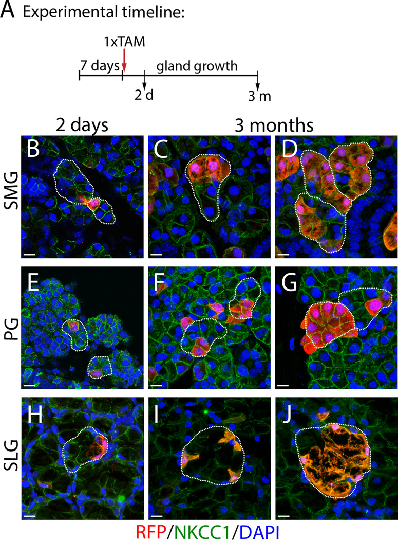
Current dogma suggests that salivary gland homeostasis is stem cell dependent. However, the extent of stem cell contribution to salivary gland maintenance has not been determined. We investigated acinar cell replacement during homeostasis, growth, and regeneration, using an inducible CreER(T2) expressed under the control of the Mist1 gene locus. Genetic labeling, followed by a chase period, showed that acinar cell replacement is not driven by the differentiation of unlabeled stem cells. Analysis using R26(Brainbow2.1) reporter revealed continued proliferation and clonal expansion of terminally differentiated acinar cells in all major salivary glands. Induced injury also demonstrated the regenerative potential of pre-labeled acinar cells. Our results support a revised model for salivary gland homeostasis based predominantly on self-duplication of acinar cells, rather than on differentiation of stem cells. The proliferative capacity of differentiated acinar cells may prove critical in the implementation of cell-based strategies to restore the salivary glands.


Conflict of interest statement

The authors declare no conflict of interest.

Figures

Figure 1
Figure 1
Acinar cells are maintained by self-duplication of pre-existing acinar cells. (A) IHC with antibody to the nuclear transcription factor, Mist1, stains acinar cells (brown) in the major salivary glands. Intercalated duct (ID); acinus is outlined. (B) Schematic diagram depicts potential outcomes of pulse-chase experiment. See text for details. (C) Experimental timeline. (D) Male and female SMGs stained for LacZ and counterstained with Nuclear Fast Red at 1 week, 2 and 6 months following tamoxifen induction. (E) Quantification of LacZ+ acinar cells per total number of acinar cells counted in glands of males and females isolated at 1week (w), and 1, 2, 3, and 6 months (m) after tamoxifen administration. See also Table S1 and Fig.S1. Scale bars= 20µm.
Figure 2
Figure 2
Clonal analysis and regeneration of acinar cells in the SMG and PG. (A) Mist1CreERT2 was crossed with the R26Brainbow2.1 reporter strain (schematic). Black triangles, LoxP sites. (B) Experimental timeline. (C) Labeled cells in the SMG at 1 week following tamoxifen treatment. (D-E) Clonal expansion of labeled cells in the SMG at 3 and 6 months after tamoxifen treatment. (F) Single labeled cells in the PG 1 week following tamoxifen treatment. (G-H) Clonal expansion of labeled cells in the PG at 3 and 6 months. (I) Model of acinar cell proliferation and clonal expansion in adult SMG and PG. (J) Quantification of clone sizes over time in adult SMG shows decrease of single cells and increase in clones of 3 cells or more (See Table S2). (K) Quantification of clone sizes over time in adult PG (See Table S2). (L) Proliferation in labeled clones in adult female SMG and PG 6 months after tamoxifen induction detected by Ki67 (red) and GFP antibody (green) (arrows). Scale bars=10 µm. (See Figs.S2, S3, Movies S1A,B). (M) Mist1CreERT2 was crossed with the R26TdTomato reporter strain (schematic). Black triangles, LoxP sites. (N) Experimental timeline. (O) Top panel: H&E staining of sections from control SMG (left) and ligated SMG (middle) after 14d ligation; and of ligated SMG (right) after 14d of regeneration. Bottom panel: RFP antibody staining (red) of control SMG (left) and ligated SMG (middle) after 14d ligation; and ligated SMG (right) after 14d of regeneration. Scale bars= 50 µm. (P) Co-localization of RFP (red) and Mist1 (green) or Nkcc1 (green) in acinar cells of control and ligated SMG after 14d regeneration. Scale bars= 20µm.
Figure 3
Figure 3
Clonal expansion of Mist1-expressing cells in the SLG. (A) Mist1CreERT2;R26LacZ mice were given tamoxifen for 4 consecutive days. LacZ expression labels serous (s) and mucous (m) acinar cells at 1 week after tamoxifen induction. (B,C) At 3 (B) and 6 months (C), LacZ positive cells were present as single cells or larger clusters. (D,G) Single labeled cells in SLG of Mist1CreERT2;R26Brainbow2.1 mice at 1 week after a single tamoxifen treatment. (E-F) After 3 and 6 months chase, some acinar cells in the adult SLG were present in monoclonal clusters (See Movie S2A). (H-I) At 3 and 6 months chase, most labelled cells in the SLG are single cells (See movie S2B). (A-C) Scale bar= 20 µm. (D-I) Scale bar= 10 µm. (J) Model of acinar cell proliferation and clonal expansion in adult SLG.
Figure 4
Figure 4
Self-duplication of acinar cells drives postnatal salivary gland expansion. (A) Experimental timeline. (B,E,H) At 9 days-old, single labeled acinar cells were found in all major salivary glands. (C,F,I) Single labeled cells or small clones in the salivary glands after 3 months chase. (D,G,J) Larger clones in all glands were also found after 3 months chase. Images show anti-RFP (red) and Nkcc1 (green) double antibody staining. Acini are indicated by outline. Scale bars= 10 µm.

References

    1. Arnold K, Sarkar A, Yram MA, Polo JM, Bronson R, Sengupta S, Seandel M, Geijsen N, Hochedlinger K. Sox2(+) adult stem and progenitor cells are important for tissue regeneration and survival of mice. Cell stem cell. 2011;9:317–329. - PMC - PubMed
    1. Bullard T, Koek L, Roztocil E, Kingsley PD, Mirels L, Ovitt CE. Ascl3 expression marks a progenitor population of both acinar and ductal cells in mouse salivary glands. Developmental biology. 2008;320:72–78. - PMC - PubMed
    1. Dardick I, Burford-Mason AP. Current status of histogenetic and morphogenetic concepts of salivary gland tumorigenesis. Critical reviews in oral biology and medicine : an official publication of the American Association of Oral Biologists. 1993;4:639–677. - PubMed
    1. Das B, Cash MN, Hand AR, Shivazad A, Culp DJ. Expression of Muc19/Smgc Gene Products During Murine Sublingual Gland Development: Cytodifferentiation and Maturation of Salivary Mucous Cells. Journal of Histochemistry & Cytochemistry. 2009;57:383–396. - PMC - PubMed
    1. Denny PC, Denny PA. Dynamics of parenchymal cell division, differentiation, and apoptosis in the young adult female mouse submandibular gland. Anatomical Record. 1999;254:408–417. - PubMed

Publication types

Substances