. 2015 Feb 12;119(6):3302-3311.
doi: 10.1021/jp512174w.

Directional Emission from Metal-Dielectric-Metal Structures: Effect of Mixed Metal Layers, Dye Location and Dielectric Thickness

Affiliations

Directional Emission from Metal-Dielectric-Metal Structures: Effect of Mixed Metal Layers, Dye Location and Dielectric Thickness

Sharmistha Dutta Choudhury et al. J Phys Chem C Nanomater Interfaces. .

Abstract

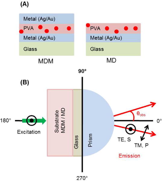
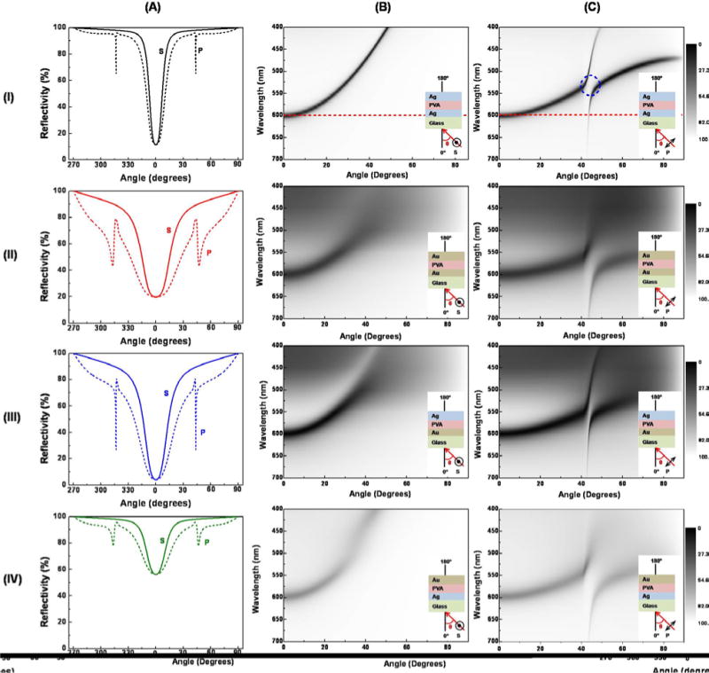
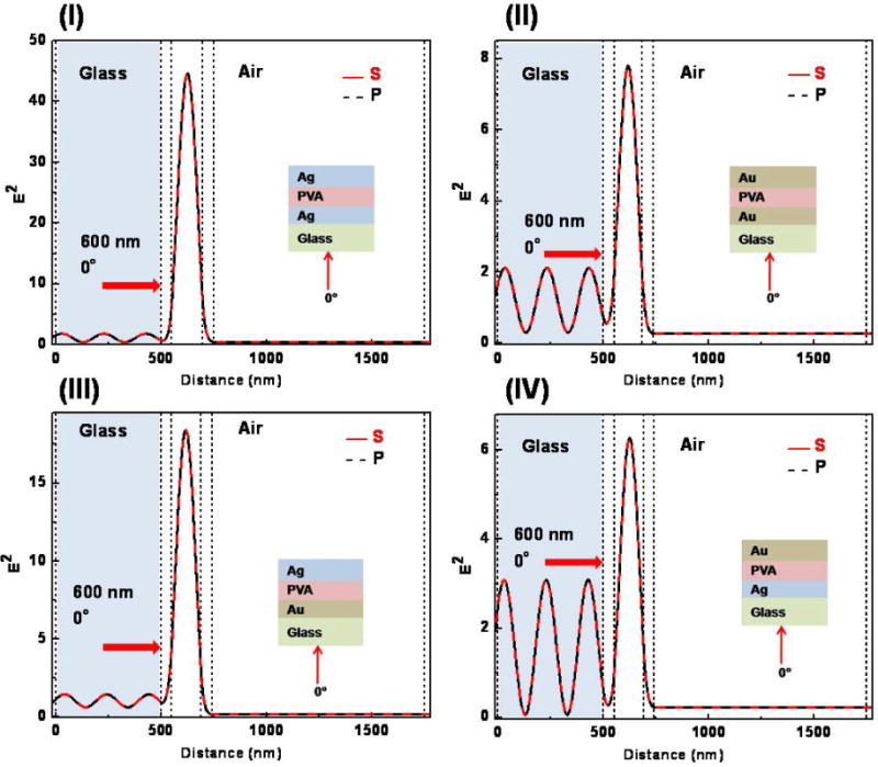
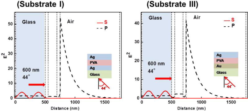
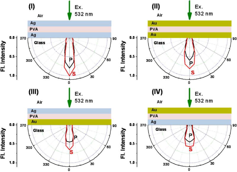
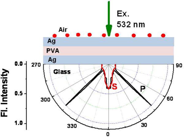
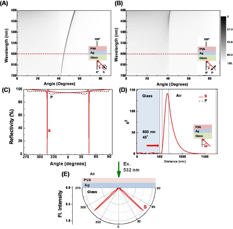
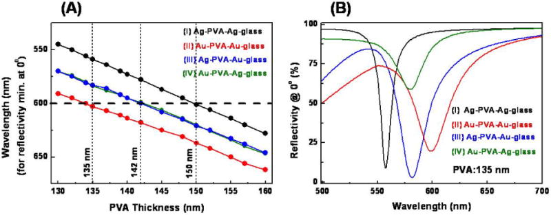
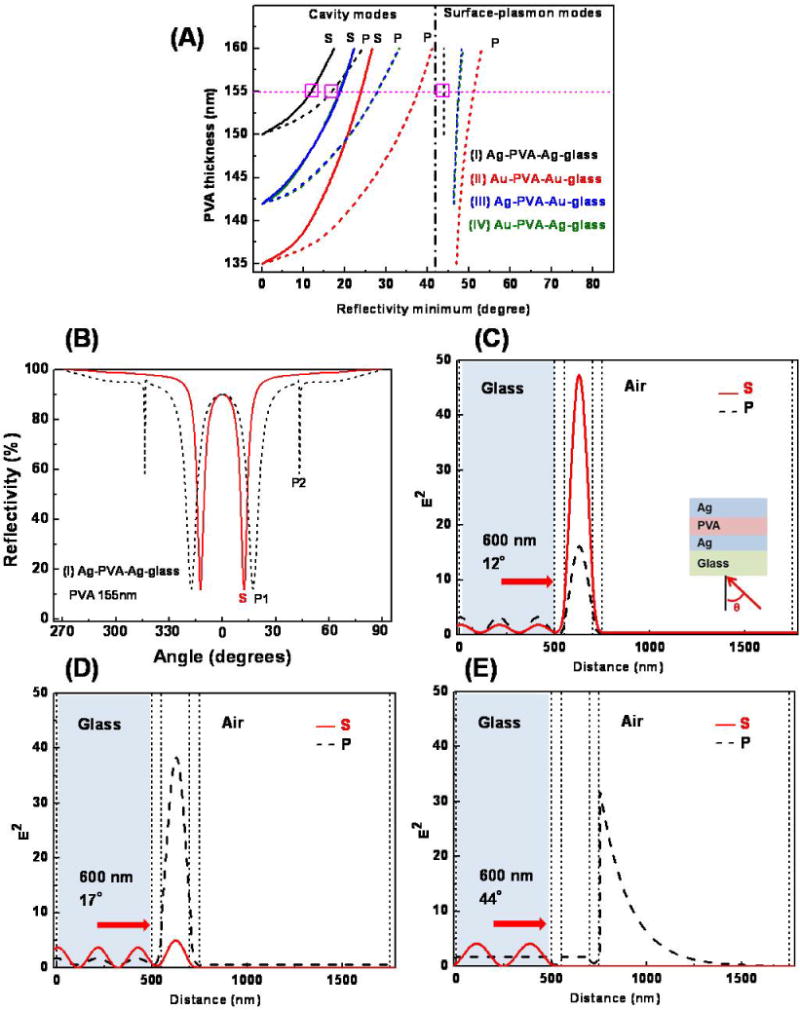
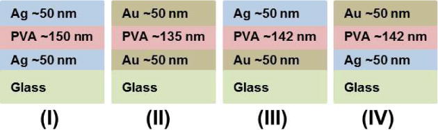
Metal-dielectric-metal (MDM) structures provide directional emission close to the surface normal, which offers opportunities for new design formats in fluorescence based applications. The directional emission arises due to near-field coupling of fluorophores with the optical modes present in the MDM substrate. Reflectivity simulations and dispersion diagrams provide a basic understanding of the mode profiles and the factors that affect the coupling efficiency and the spatial distribution of the coupled emission. This work reveals that the composition of the metal layers, the location of the dye in the MDM substrate and the dielectric thickness are important parameters that can be chosen to tune the color of the emission wavelength, the angle of observation, the angular divergence of the emission and the polarization of the emitted light. These features are valuable for displays and optical signage.

Keywords: Cavity-Mode-Coupled Emission; Directional Emission; Dispersion; Metal-Dielectric-Metal; Surface-Plasmon-Coupled Emission.


Figures

Figure 1
Figure 1
(A) Schematic of the metal-dielectric-metal (MDM) and metal-dielectric (MD) substrates. The dielectric (PVA) layer contains the dye molecules, S101. (B) Experimental geometry and polarizations used for the present measurements; the excitation laser (532 nm) is vertically polarized, which is normal to the plane of the paper.
Figure 2
Figure 2
(A) Calculated angle-dependent reflectivity for MDM substrates (I–IV) with 600 nm incident light. (B) Reflectivity dispersion diagrams, R(λ, θ), for S-polarized illumination for substrates (I–IV). (C) Reflectivity dispersion diagrams for P-polarized illumination for substrates (I–IV); reflectivity scale from 0 to 100%. Insets in B and C show the MDM structure, illumination geometry and polarization. PVA thickness for substrate I: 150 nm, Substrate II: 135 nm, Substrates III and IV: 142 nm.
Figure 3
Figure 3
Calculated electric field intensities in different MDM substrates (I–IV) for 600 nm illumination at 0°; S-polarized (red) and P-polarized (black).
Figure 4
Figure 4
Calculated electric field intensities in MDM substrate I and III for 600 nm illumination at 44°; S-polarized (red) and P-polarized (black). Corresponding electric field distributions for the substrates II and IV are shown in Figure S1, SI.
Figure 5
Figure 5
Angle-dependent S- (red) and P-polarized (black) emission intensities at 600 nm from S101 embedded in the PVA layer in MDM substrates (I–IV). The intensities have been normalized with respect to the maximum intensity observed for substrate I.
Figure 6
Figure 6
Angle-dependent S- (red) and P-polarized (black) emission intensities at 600 nm from S101 placed on the top metal layer in the MDM substrate I.
Figure 7
Figure 7
Calculated reflectivity dispersion diagrams, R(λ, θ), for (A) S-polarized and (B) P-polarized illumination in the PVA-Ag-glass MD substrate; (C) angle-dependent reflectivity with 600 nm incident light; (D) electric field intensity for 600 nm illumination (S- and P-polarized) at the reflectivity minimum of 45° and (E) experimentally observed angle-dependent emission intensities at 600 nm from S101 embedded in the PVA layer, PVA thickness 150 nm.
Figure 8
Figure 8
(A) Variation of the beaming wavelength (observed normal to the substrate at 0°) with PVA thickness in different MDM substrates. The wavelength of interest in the present study (600 nm) is shown by a dashed horizontal line. (B) Wavelengths at which the reflectivity dip at 0° is obtained in different MDM substrates for a particular PVA thickness (135 nm).
Figure 9
Figure 9
(A) Angular dependence of the reflectivity minima on the PVA thickness in MDM substrates I–IV, plots for substrate III and IV are similar and superimposed. (B) Angle-dependent reflectivity for Ag-PVA-Ag-glass MDM substrate with PVA thickness of 155 nm. (C–E) Electric field intensities in the Ag-PVA-Ag-glass substrate with PVA thickness of 155 nm for each resonance angle (shown by horizontal line in A). All calculations have been performed for 600 nm (S- and P-polarized).
Scheme 1
Scheme 1
Schematic of the metal-dielectric-metal (MDM) substrates used in the present study.

Similar articles

Cited by

References

    1. Lakowicz JR, Shena Y, D’Auria S, Malicka J, Fang J, Gryczynski Z, Gryczynski I. Radiative decay engineering: 2. Effects of silver island films on fluorescence intensity, lifetimes, and resonance energy transfer. Anal Biochem. 2002;301:261–267. - PMC - PubMed
    1. Lakowicz JR. Radiative Decay Engineering 3. Surface Plasmon-Coupled Directional Emission. Anal Biochem. 2004;324:153–169. - PMC - PubMed
    1. Gryczynski I, Malicka J, Gryczynski Z, Lakowicz JR. Radiative Decay Engineering 4. Experimental Studies of Surface Plasmon-Coupled Directional Emission. Anal Biochem. 2004;324:170–182. - PMC - PubMed
    1. Gryczynski I, Malicka J, Gryczynski Z, Lakowicz JR. Surface Plasmon-Coupled Emission with Gold Films. J Phys Chem B. 2004;108:12568–12574. - PMC - PubMed
    1. Enderlein J, Ruckstuhl T. The Efficiency of Surface-Plasmon Coupled Emission for Sensitive Fluorescence Detection. Optics Express. 2005;13:8855–8865. - PubMed

LinkOut - more resources