. 2015 Apr 6;5(2):20140078.
doi: 10.1098/rsfs.2014.0078.

Multiscale modelling of the feto-placental vasculature

Affiliations

Multiscale modelling of the feto-placental vasculature

A R Clark et al. Interface Focus. .

Abstract

The placenta provides all the nutrients required for the fetus through pregnancy. It develops dynamically, and, to avoid rejection of the fetus, there is no mixing of fetal and maternal blood; rather, the branched placental villi 'bathe' in blood supplied from the uterine arteries. Within the villi, the feto-placental vasculature also develops a complex branching structure in order to maximize exchange between the placental and maternal circulations. To understand the development of the placenta, we must translate functional information across spatial scales including the interaction between macro- and micro-scale haemodynamics and account for the effects of a dynamically and rapidly changing structure through the time course of pregnancy. Here, we present steps towards an anatomically based and multiscale approach to modelling the feto-placental circulation. We assess the effect of the location of cord insertion on feto-placental blood flow resistance and flow heterogeneity and show that, although cord insertion does not appear to directly influence feto-placental resistance, the heterogeneity of flow in the placenta is predicted to increase from a 19.4% coefficient of variation with central cord insertion to 23.3% when the cord is inserted 2 cm from the edge of the placenta. Model geometries with spheroidal and ellipsoidal shapes, but the same volume, showed no significant differences in flow resistance or heterogeneity, implying that normal asymmetry in shape does not affect placental efficiency. However, the size and number of small capillary vessels is predicted to have a large effect on feto-placental resistance and flow heterogeneity. Using this new model as an example, we highlight the importance of taking an integrated multi-disciplinary and multiscale approach to understand development of the placenta.

Keywords: computational model; feto–placental circulation; multiscale; placenta; pregnancy; vascular structure.


Figures

Figure 1.
Figure 1.
A schematic of the structure of the placental vasculature. Deoxygenated blood is delivered to the placenta via two umbilical arteries. These arteries branch into large chorionic vessels which feed 60–100 individual villous trees. The villous trees themselves branch dichotomously for several generations before feeding several parallel capillary conduit pathways, which provide the site for placental gas exchange. Several parallel pathways between arteries and veins in the villous tree are incorporated at the level of the intermediate and terminal villi in the model developed here, as have been observed in cast- and imaging-based studies. However, for simplicity, only a single artery and vein per villous branch are shown in the figure (inset). (Online version in colour.)
Figure 2.
Figure 2.
(a) Schematic of the area-filling branching algorithm of Wang et al. [38]. Initial placement of the umbilical arteries is assigned and seed points are split into two groups. (a) The centre of mass of seed points in each group is calculated and a new artery is grown a prescribed distance towards this centre of mass. (b) The collection of seed points is split according to the centrelines of the newly generated arteries, centres of mass for each subregion are recalculated and four new branches generated a fixed distance towards these points. The process is repeated until there is only one seed point left in each group. (Online version in colour.)
Figure 3.
Figure 3.
Graphical depictions of the model geometry. (a) An intraplacental vessel which is much smaller in diameter than the main chorionic vessels arising from a chorionic vessel at close to right angles. The largest placental arteries in the case of (b) central and (c) non-central cord insertion. In accordance with previous studies the case with central cord insertion is relatively more symmetric than the case of non-central insertion. Veins and peripheral small arteries and capillaries are not shown. (Online version in colour.)
Figure 4.
Figure 4.
Illustrations of the model description of terminal capillary conduits. (a) In the placenta mature intermediate villus vessels branch and each intermediate villus gives rise to on average six terminal convolutes that follow tortuous pathways from artery to vein. (b) We model these terminal convolutes as arising consecutively from intermediate villus arteries and neglect any connections between capillary convolutes. (c) Finally, we assume that intermediate villus arteries branch symmetrically so the number of vessels doubles at each generation. Resistance of the terminal portions of the placental vasculature can then be summed in serial and parallel through the system. (Online version in colour.)
Figure 5.
Figure 5.
Model simulations of the effective resistance of a terminal unit, expressed as the effective length of a single vessel with the same radius as an intermediate villus, plotted against the number of parallel capillary connections associated with an intermediate villus. The model suggests that parallel capillary connections contribute to maximizing the capillary surface area available to gas exchange while allowing the resistance of the placental vasculature to stay low.
Figure 6.
Figure 6.
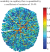
An illustration of model predictions of the heterogeneous distribution of flow to placental capillaries. Each capillary unit is represented as a sphere and the relative flow in each sphere is represented by a linear colour scale. Large placental arteries are also shown, coloured by predicted blood pressure. (Online version in colour.)
Figure 7.
Figure 7.
Model predictions of the umbilical artery pressure as a function of the number of parallel capillary connections associated with an intermediate villus. This indicates that, in pathological pregnancies with insufficient parallel capillary connections, an increased umbilical artery pressure could put additional strain on the fetal heart.
Figure 8.
Figure 8.
Model predictions on the effect of intermediate villous artery and capillary convolute calibre on feto–placental resistance. This effect is highly nonlinear and a halving on capillary convolute radius has a significant impact on predicted resistance. Capillary convolute radius has the greatest effect on the resistance of the system as a whole as the placental capillaries comprise a large number of small vessels.

References

    1. Teasdale F, Jean-Jaques G. 1988. Intrauterine growth retardation: morphometry of the microvillous membrane of the human placenta. Placenta 9, 47–55. (10.1016/0143-4004(88)90072-0) - DOI - PubMed
    1. Kingdom J, Huppertz B, Seaward G, Kaufmann P. 2000. Development of the placental villous tree and its consequences for fetal growth. Eur. J. Obstet. Gynecol. Reprod. Biol. 92, 35–43. (10.1016/S0301-2115(00)00423-1) - DOI - PubMed
    1. Jirkovská M, Kubínová L, Krekule I, Hach P. 1998. Spatial arrangement of fetal placental capillaries in terminal villi: a study using confocal microscopy. Anat. Embryol. 197, 263–272. (10.1007/s004290050136) - DOI - PubMed
    1. Gordon Z, Elad D, Almog R, Hazan Y, Jaffa A, Eytan O. 2007. Anthropometry of fetal vasculature in the chorionic plate. J. Anat. 211, 698–706. (10.1111/j.1469-7580.2007.00819.x) - DOI - PMC - PubMed
    1. Benirschke K, Kaufmann P. 1990. Pathology of the human placenta, pp. 180–243. New York, NY: Springer.