. 2015 Mar 1;2(1):47-57.
doi: 10.1093/rb/rbu016.

Functionalized scaffolds to enhance tissue regeneration

Affiliations

Functionalized scaffolds to enhance tissue regeneration

Baolin Guo et al. Regen Biomater. .

Abstract

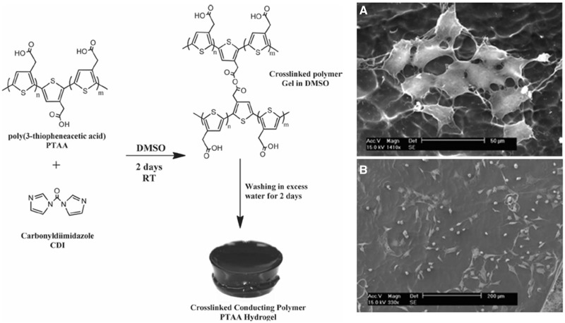
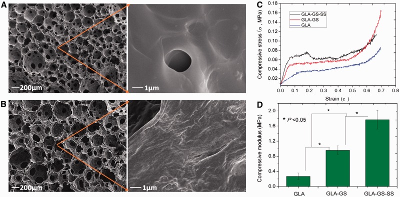
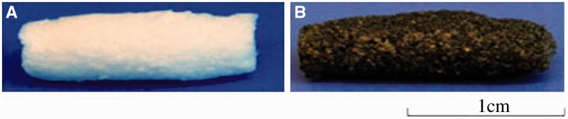
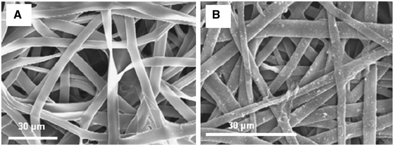
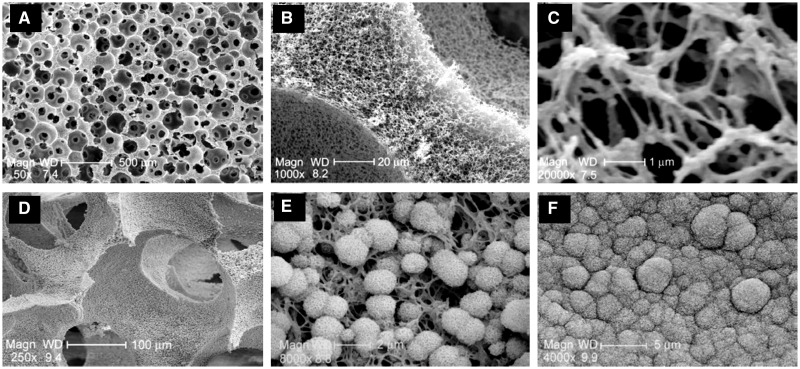
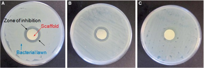
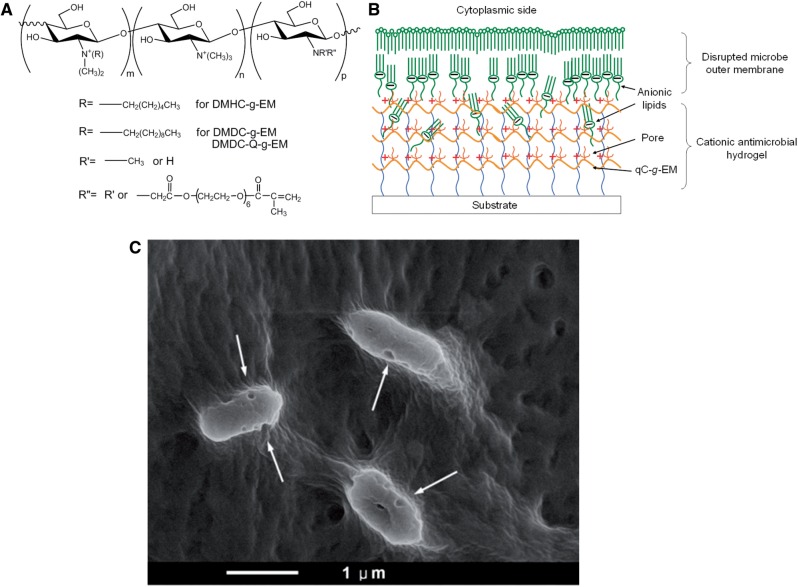
Tissue engineering scaffolds play a vital role in regenerative medicine. It not only provides a temporary 3-dimensional support during tissue repair, but also regulates the cell behavior, such as cell adhesion, proliferation and differentiation. In this review, we summarize the development and trends of functional scaffolding biomaterials including electrically conducting hydrogels and nano-composites of hydroxyapatite (HA) and bioactive glasses (BGs) with various biodegradable polymers. Furthermore, the progress on the fabrication of biomimetic nanofibrous scaffolds from conducting polymers and composites of HA and BG via electrospinning, deposition and thermally induced phase separation is discussed. Moreover, bioactive molecules and surface properties of scaffolds are very important during tissue repair. Bioactive molecule-releasing scaffolds and antimicrobial surface coatings for biomedical implants and scaffolds are also reviewed.

Keywords: antimicrobial coatings; bioactive nanocomposites; biomaterials; bone tissue engineering; electrically conductive polymers; molecule-releasing scaffolds; scaffolds.


Figures

Figure 1.
Figure 1.
Schematic preparation of conductive hydrogel (left column). Representative SEM images of myoblast cells adhered to the conductive hydrogel substrates after 72 h incubation (right column) (A: high magnification, and B: low magnification) [19]. Copyright 2012. With the permission of Wiley.
Figure 2.
Figure 2.
Typical gelatin–silica BG hybrid scaffolds prepared by direct foaming–freezing method. The molecular-level distributions in polymer matrix and significantly improved mechanical properties can be obtained after direct hybridization process. (A-B) Scaffolds morphology of pure gelatin (A) and gelatin-BG hybrids; (C-D) Stress-strain curves (C) and compressive strength (D) of samples (GLA: gelatin; GS: siloxane; SS: silica-based glass). Reproduced from Ref. [25] with permission from Elsevier.
Figure 3.
Figure 3.
Photographs of the degradable tubular porous scaffolds from PCL (A) and degradable conductive porous scaffolds from blends of PCL/hyperbranched degradable conducting polymer (B) [53]. Copyright 2012. With the permission of Elsevier.
Figure 4.
Figure 4.
SEM images of the nanofibers. (A) Superficial aspect of non-coated SF mesh and (B) SF-PPY-coated mesh [66]. Copyright 2012. With the permission of Elsevier.
Figure 5.
Figure 5.
Biomimetic gelatin and gelatin–apatite nanofibrous scaffolds fabricated by TIPS and biomineralization methods. (A–C) Morphology and microstructure of nanofibrous scaffolds; (D–F) scaffolds after biomineralization for 7 days (D and E) and 21 days (F). Reproduced from Ref. [31] with permission from Elsevier.
Figure 6.
Figure 6.
Functionalized PLA nano-fibrous scaffolds incorporating recombinant human bone morphogenetic protein-7 (rhBMP-7) nanospheres. (A, B) rhBMP-7-loaded nanosphere and scaffolds; (C–F) SEM images of nano-fibrous scaffolds before (C and D) and after nanosphere incorporation (E and F). Reproduced from Ref. [31] with permission from Elsevier.
Figure 7.
Figure 7.
Antimicrobial properties of PLA nanofibrous scaffolds treated with Silvadur ET containing 31.25 μg/ml silver against Escherichia coli (A), Staphylococcus aureus (B) and silver-resistant E. coli (C) bacteria as evaluated by the AATCC 147 test [92]. Copyright 2014. With the permission of Elsevier.
Figure 8.
Figure 8.
Nanoporous antimicroibial hydrogel coating fabricated by polysaccharides: (A) synthesis of quaternized CS functionalized with acrylate PEG side-chains; (B) formation of nanoporous hydrogel coating which is capable to kill microbes; (C) the cell wall of the Gram-negative bacteria Pseudomonas aeruginosa was disrupted by the nanoporous hydrogel. Reproduced from Ref. [105] with permission from Nature Publishing Group.

References

    1. Chen GP, Ushida T, Tateishi T. Scaffold design for tissue engineering. Macromol Biosci 2002;2:67–77.
    1. Hubbell JA. Biomaterials in tissue engineering. Bio-Technology 1995;13:565–76. - PubMed
    1. Liu XH, Ma PX. Polymeric scaffolds for bone tissue engineering. Ann Biomed Eng 2004;32:477–86. - PubMed
    1. Ma PX. Biomimetic materials for tissue engineering. Adv Drug Delivery Rev 2008;60:184–98. - PMC - PubMed
    1. Guo BL, Ma PX. Synthetic biodegradable functional polymers for tissue engineering—a brief review. Sci China Chem 2014;57:490–500. - PMC - PubMed

LinkOut - more resources