. 2015 Feb 19;3(3):1-36.
doi: 10.1128/microbiolspec.MDNA3-0047-2014.

Site-specific DNA Inversion by Serine Recombinases

Affiliations

Site-specific DNA Inversion by Serine Recombinases

Reid C Johnson. Microbiol Spectr. .

Abstract

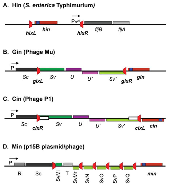
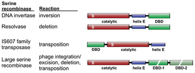
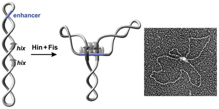
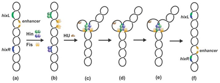
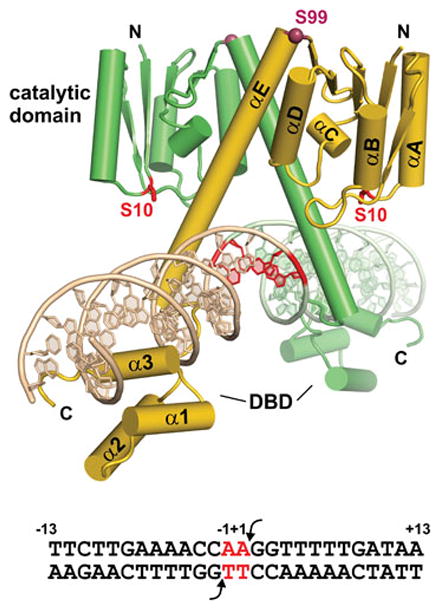
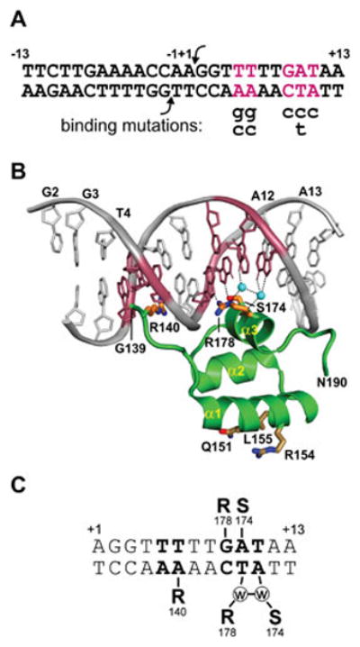
Reversible site-specific DNA inversion reactions are widely distributed in bacteria and their viruses. They control a range of biological reactions that most often involve alterations of molecules on the surface of cells or phage. These programmed DNA rearrangements usually occur at a low frequency, thereby preadapting a small subset of the population to a change in environmental conditions, or in the case of phages, an expanded host range. A dedicated recombinase, sometimes with the aid of additional regulatory or DNA architectural proteins, catalyzes the inversion of DNA. RecA or other components of the general recombination-repair machinery are not involved. This chapter discusses site-specific DNA inversion reactions mediated by the serine recombinase family of enzymes and focuses on the extensively studied serine DNA invertases that are stringently controlled by the Fis-bound enhancer regulatory system. The first section summarizes biological features and general properties of inversion reactions by the Fis/enhancer-dependent serine invertases and the recently described serine DNA invertases in Bacteroides. Mechanistic studies of reactions catalyzed by the Hin and Gin invertases are then discussed in more depth, particularly with regards to recent advances in our understanding of the function of the Fis/enhancer regulatory system, the assembly of the active recombination complex (invertasome) containing the Fis/enhancer, and the process of DNA strand exchange by rotation of synapsed subunit pairs within the invertasome. The role of DNA topological forces that function in concert with the Fis/enhancer controlling element in specifying the overwhelming bias for DNA inversion over deletion and intermolecular recombination is emphasized.


Figures

Figure 1
Figure 1
Genetic organization of Fis/enhancer-dependent DNA inversion systems. A. The Hin system controlling flagellin synthesis in Salmonella enterica Typhimurium. fljB codes for one of the alternatively expressed flagellins, and fljA is a repressor of the fliC flagellin gene located elsewhere on the chromosome. B and C. The Gin and Cin systems from phages Mu and P1, which control phage host range. Sv and Sv′ gene segments are alternatively fused in-frame to the Sc gene segment. The S and U genes encode tail fiber proteins. D. The complex Min locus from the p15B plasmid. The different Sv gene segments are alternatively fused to the Sc gene segment. In each case the recombination sites are colored red, and the positions of the DNA invertases (maroon) with their associated recombinational enhancer segments (blue) are denoted. P designates a promoter with the S. Typhimurium fliC and fljA genes being transcribed by the sigma 28 form of RNA polymerase.
Figure 2
Figure 2
Fis/enhancer-dependent serine DNA invertase recombination sites. DNA sequences of recombination sites from the systems depicted in Fig. 1 are listed along with the consensus sequence at the top. Sequences matching the consensus are highlighted in cyan.
Figure 3
Figure 3
Amino acid sequence alignment of the Hin and Gin DNA invertases together with γδ resolvase. Highly conserved residues among all serine recombinases are highlighted in magenta. The active site serine residue is shaded black. Highly conserved residues among Fis/enhancer-dependent DNA invertases are in cyan; many of these are conserved among the small serine recombinases. Yellow highlighted residues within the α-helix B region are those that contact enhancer DNA and within α-helix 1 of the DNA binding domain (Hin residues 139–190) are those that interact with Fis. Residue numbering is according to Hin. Asterisks denote residues where single substitutions result in strong gain-of-function activities, often resulting in Fis/enhancer-independence (65, 109, 185). Secondary structure designations are from γδ resolvase (residues 1–138; PDB: 1GDT) and Hin (residues 139–190; PDB: 1IJW). An alignment that includes additional Fis/enhancer-dependent DNA invertases along with other small serine recombinases is given in ref. (2).
Figure 4
Figure 4
Serine recombinase subfamilies. The domain architectures of serine recombinase subfamily members denoting the ~100 amino acid residue catalytic domain containing the active site serine (S), the oligomerization helix E, and the DNA binding domain (DBD). The DNA binding regions of large serine recombinases can be quite large (300–450 residues) and consist of two discrete domains. The DNA invertases and resolvases are often grouped together as small serine recombinases.
Figure 5
Figure 5
The Hin invertasome. Electron micrograph of an invertasome together with a schematic drawing of the structure. The invertasome structure was stabilized by crosslinking and supercoils removed prior to spreading onto the grid and low angle platinum shadowing (104). Hin subunits are rendered as translucent spheres; Fis subunits are rendered as ovals. Hix sites are depicted as arrows and the enhancer segment is blue.
Figure 6
Figure 6
DNA inversion reaction pathway by Fis/enhancer-dependent serine invertases. In step (c) Hin dimers bound to hixL and hixR are associated with the Fis-bound enhancer at the base of a branch on supercoiled DNA. Formation of the Hin tetramer (c) generates an enzyme active for double strand cleavage and subunit rotation (d). Ligation and resolution of the complex (e) results in inversion of the DNA segment between recombination sites.
Figure 7
Figure 7
The serine DNA invertase dimer. Model of the Hin dimer bound to hixL derived from the catalytic domain and oligomerization helix E of γδ resolvase (PDB: 1GDT) linked to the DNA binding domain (DBD) of Hin (PDB: 1IJW) is shown. The Ser10 active site residue and core nucleotides where DNA exchange occurs are colored red. Hinge residue Ser99 (Cα) is rendered as a dark red sphere. The sequence of hixL showing the Hin cleavage sites (arrows) and core nucleotides (red) is given below.
Figure 8
Figure 8
The Hin DNA binding domain bound to the conserved hix half-site located within the invertible segment. A. Sequence of hixL with the locations of severe DNA binding mutations (99, 123). B. Hin DBD – DNA complex (PDB: 1IJW). Side chains of residues Arg140, Ser174, and Arg178 along with the two ordered water molecules (cyan spheres) that make critical base contacts are shown. Conserved residues Gln151, Arg154, and Leu155 on α-helix 1 that contact Fis are also shown. C. Schematic representation of sequence-specific contacts, including water bridged hydrogen bonding.
Figure 9
Figure 9
Fis and the recombinational enhancer. A. Fis binding motif derived from footprinting, mutagenesis, genome-wide ChIP, and X-ray crystallography (see ref. (130)). Bases below the numbering are strongly inhibitory for binding. B. Structure of the Fis dimer bound to a high affinity DNA segment (Fis residues 10–98; PDB: 3IV5); the sequence of the 15 bp core between ±7 (colored brown) is given below. Arg85 contacts the conserved guanines at the borders of the core sequence, Asn84 contacts the DNA backbone and often the base at ±4 and is responsible for the inhibitory effect of a thymine at this position (panel A). A subset of other important residues making DNA backbone contacts are colored grey. The Arg71 side chains, which are poorly resolved in most structures of DNA complexes, are shown oriented towards DNA. Bending of the flanking DNA segments varies depending on the DNA sequence. The triad of residues (Val16, Asp20, and Val22) near the tips of the mobile β-hairpin arms that contact DNA invertases are denoted for the cyan colored subunit. (C) Model of the hin enhancer. The two Fis dimers are docked onto the hin enhancer DNA sequence. The Fis β-hairpin arms are highlighted in red. The A/T-rich DNA segments contacted by the helix B regions of the DNA invertase tetramer in the invertasome are colored magenta (78).
Figure 10
Figure 10
Assembly of the Hin invertasome and subunit rotation. A. Hix-Hin dimers associated at the Fis-bound enhancer. Fis dimers are gold with their β-hairpin arms colored magenta. Hin α-helix B and α-helix 1 are colored red. B. Pre-activated Hin tetramer (based on 3BVP) and C. post-cleavage tetramer (based on 1ZR4). Side chains of residues from helix B that contact enhancer DNA are denoted. D and E. Partial rotations (50° and 90°, respectively) of the top synaptic subunit pair and (F) complete subunit rotation to mediate the exchange of DNA strands. A movie depicting the assembly of the invertasome and DNA exchange by subunit rotation is provided in Video 2 of reference (78), from which these images are taken. Details of the models are described in ref. (78, 109, 117).
Figure 11
Figure 11
DNA topological changes during the inversion reaction. The starting complex (invertasome) between the two recombination sites (arrows) and the enhancer at the base of a plectonemic branch traps two negative DNA nodes (DNA depicted as a ribbon without supercoiling). Double strand cleavages and DNA exchange by the equivalent of a 180° clockwise rotation create a negative node but also introduce two half turns of helical twist that cancel the negative node. The recombinant configuration of DNA strands changes the trapped nodes to a positive sign resulting in an overall linking number change of +4. Node signs are determined by directionally tracing the entire path of the DNA molecule (193). By convention, a node is defined as negative when the DNA strand in front is pointed upwards and the strand underneath crosses in a rightward direction. A positive node is when the strand underneath crosses in a leftward direction. (Figure is modified from ref. (105)).
Figure 12
Figure 12
DNA topological changes during processive recombination by serine DNA invertases. Initially, the recombination sites (green and blue) are assembled at the enhancer DNA (brown) in the invertasome structure. The initial assembly depicts short and long loops between the recombination sites and enhancer as found in the Hin and Gin systems. The first DNA exchange by the equivalent of 180° rotations of DNA strands covalently-linked to recombinase subunits generates inversion; these molecules have lost 4 negative supercoils (not shown) but no knot nodes are introduced. Subsequent processive rounds of DNA exchange generate knots of increasing complexity with the orientation of the invertible segment alternating between parental and inverted; these have all lost 2 negative supercoils. Multiple windings of DNA (processive DNA exchanges) are torsionally restricted as long as the enhancer remains associated with the recombination sites, but release of the enhancer removes this constraint. The structures of the knots, which have been confirmed by electron microscopy, are depicted below.
Figure 13
Figure 13
Synapsis and DNA exchange of recombination sites. A. Synapsis of wild-type recombination sites in a parallel configuration as in the standard invertasome complex. DNA cleavage followed by exchange and ligation inverts the intervening DNA segment. B. Synapsis where one recombination site contains a mutation (red) within the core nucleotides. DNA cleavage and a single exchange result in unpaired core nucleotides that cannot ligate. A second exchange is required for ligation, which reorients the intervening DNA segment into the starting (parental) configuration and generates a 3-noded knot (Fig. 12). C. Synapsis of wild-type recombination sites that are oriented in a directly-repeated configuration. Most of the time the sites align in an antiparallel configuration within an invertasome structure (see Fig. 16), and the core nucleotides cannot base pair after the first DNA exchange. Ligation can occur after a second exchange back into the parental orientation creating a 3-noded knot as in panel B.
Figure 14
Figure 14
Site-directed crosslinking demonstrating subunit rotation within active Hin tetramers. Model of the Hin tetramer based on resolvase tetramer structures. View is looking into the rotation interface with residues converted to cysteine for site-directed crosslinking rendered as colored spheres (Cβ atom). Rotations refer to the top subunit pair (gold and purple) relative to a fixed bottom subunit pair. M101C residues (orange spheres) from the gold and blue trans-diagonally located subunits are within range for crosslinking with a bis-thio reactive reagent containing an 8 Å spacer but becomes optimally positioned for crosslinking after a ~20° clockwise (cw) rotation. Initial crosslinks at residue 101 are between gold and blue subunits, but with time, purple and blue crosslinked subunit products that are the result of subunit exchange become equally represented. Crosslinking between S94C residues (magenta spheres) requires either a counterclockwise (ccw) rotation of at least 20° (optimal between 40–70°) to link trans-diagonally subunits or a clockwise rotation of about 270° to link purple and blue subunits from an original dimer. Fis/enhancer-activated reactions on supercoiled DNA primarily form 94–94 crosslinks between purple and blue subunits (cw rotation), whereas crosslinks between gold and blue subunits (ccw rotation) are overrepresented initially in Fis/enhancer-independent reactions but then both products become equally represented. Red spheres mark residues within the C-terminal third of helix E that support robust crosslinking even though they can be up to 70 Å from their nearest partner; rotations of 90 ± 15° in the cw or ccw direction are required to generate crosslinks. Fis/enhancer-activated reactions on supercoiled DNA primarily form crosslinks between purple and green subunits (cw rotation) containing helix E cysteines, whereas Fis/enhancer-independent reactions without DNA supercoiling form crosslinks between gold and green subunits (ccw) or purple and green subunits (cw) with the same kinetics. Dashed lines highlight the positions of Q134C residues at the C-terminal ends of helix E. The kinetics of crosslinking between residues at positions 101, 94, and the C-terminal end of helix E provide strong support for bidirectional subunit rotation within Fis/enhancer-independent reactions and for primarily single round cw rotation in Fis/enhancer-dependent reactions on negatively supercoiled DNA. Crosslinks between helix D residues like K72C (green spheres) readily form upon full assembly of the tetramer and do not block subunit rotation or DNA ligation. Details are provided in refs. (117, 118, 158).
Figure 15
Figure 15
The subunit rotation interface. Surfaces of rotating subunit pairs from the (A) Hin model (residues 2–134 based on PDB: 2GM4), (B) Gin X-ray structure (residues 2–125, PDB: 3UJ3), and (C) γδ resolvase X-ray structure (residues 1–132, PDB: 2GM4) are shown after alignment. Hydrophobic residues are colored yellow, acidic residues are red, basic residues are blue, and polar residues are green. A 1.6 Å probe was used to render the surfaces. Dashed circles demarking the rotating interface have a diameter of ~20 Å. D. Surface area overlap calculated for different clockwise rotational conformers from the Hin model; γδ resolvase gives a very similar pattern (117, 119). Rotations of around 0–10° correspond to conformers where the DNA ends are in-line for ligation and conformers around 100° have the E-helices between dimer pairs in a parallel/antiparallel configuration. The Hin models are based on γδ resolvase structures (shown here based on 2GM4); comparison of subunit structures with those from resolvase tetramers (2GM4 or 1ZR4) give RMSD values of <0.7 Å over the peptide backbone (residues 1–120). Subunits from the Gin tetramer structure (3UJ3) exhibit RMSD values of 1.3 – 1.5 Å over the peptide backbone atoms from residues 3–120 and 1.1 – 1.4 Å over just the catalytic domains from the Hin models or γδ resolvase tetramers (1ZR4 or 2GM4). Much of the difference between Gin and resolvase structures or Hin models is over poorly resolved loops connecting β1 to αA and β2 to αB.
Figure 16
Figure 16
Products formed on substrates containing directly-repeated recombination sites. Most synaptic complexes assemble in an invertasome structure as diagrammed in the top pathway, but the recombination sites are in an antiparallel orientation. The unpaired core nucleotides after a single DNA exchange prevent ligation (Fig. 13C). Ligation after two exchanges results in a knot containing three negative nodes (Fig. 12), as shown in the electron micrograph of a trefoil generated by Hin. In the bottom pathway, which occurs rarely, the recombination sites assemble in parallel orientation that requires an additional DNA loop. This structure is energetically unfavorable on a negatively supercoiled substrate. A single DNA exchange results in a singly-linked catenated deletion product, as shown in the micrograph. The DNA molecules were coated with RecA protein to facilitate visualization of the DNA nodes.
Figure 17
Figure 17
Residues regulating conformational transitions. A. Hin dimer model depicting some of the residues where mutations have been isolated that control the dimer-tetramer transition. B. Hin tetramer model (based on PDB: 2GM4) highlighting helix E residues Phe105 and Met109, whose side chains are located within the rotation interface and His107, whose side chain is predicted to stabilize the synaptic interface. C. Subunit from the Hin dimer model showing helix E residues Phe104, Phe105, Phe106 (behind helix E), and Met109. The surface of the catalytic domain is also shown highlighting the pocket organized by residues surrounding Phe88. Residues around the Phe88 pocket where mutations lead to Fis/enhancer-independence are colored red. D. Subunit from the Hin tetramer model rendered similarly as in panel C. Note that the side chains of Phe105 and Met109 have rotated away from the catalytic domain and are now within the rotation interface (panel B). Phe104 rotates away from the partner dimer subunit and becomes associated with the synaptic and rotation interfaces. Certain mutations at His87 (colored maroon) lead to strong Fis/enhancer-independence, and different substitutions of the hinge residue Ser99 generate various phenotypes (see text). E. Subunit from the Gin tetramer X-ray structure (PDB: 3UJ3) in the reverse orientation as shown in panel D. Numbering of residues in Gin is one less than Hin.

Similar articles

Cited by

References

    1. Grindley ND, Whiteson KL, Rice PA. Mechanisms of site-specific recombination. Annu Rev Biochem. 2006;75:567–605. - PubMed
    1. Johnson RC. Bacterial site-specific DNA inversion systems. In: Craig NL, Craigie R, Gellert M, Lambowitz AM, editors. Mobile DNA II. ASM Press; Washington, D.C: 2002. pp. 230–271.
    1. Rice PA. Title to be added. In: Craig NL, editor. Mobile DNA III. ASM Press; Washington, D.C: 2014. in press.
    1. Stark WM. The serine recombinases. In: Craig NL, editor. Mobile DNA III. ASM Press; Washington, D.C: 2014. in press.
    1. Andrewes FW. Studies in group-agglutination - The Salmonella group and its antigenic structure. J Pathology and Bacteriology. 1922;25:505–521.

LinkOut - more resources