

Co-localization between the BOLD response and epileptiform discharges recorded by simultaneous intracranial EEG-fMRI at 3 T
- PMID: 25844327
- PMCID: PMC4375646
- DOI: 10.1016/j.nicl.2015.03.002
Co-localization between the BOLD response and epileptiform discharges recorded by simultaneous intracranial EEG-fMRI at 3 T
Abstract
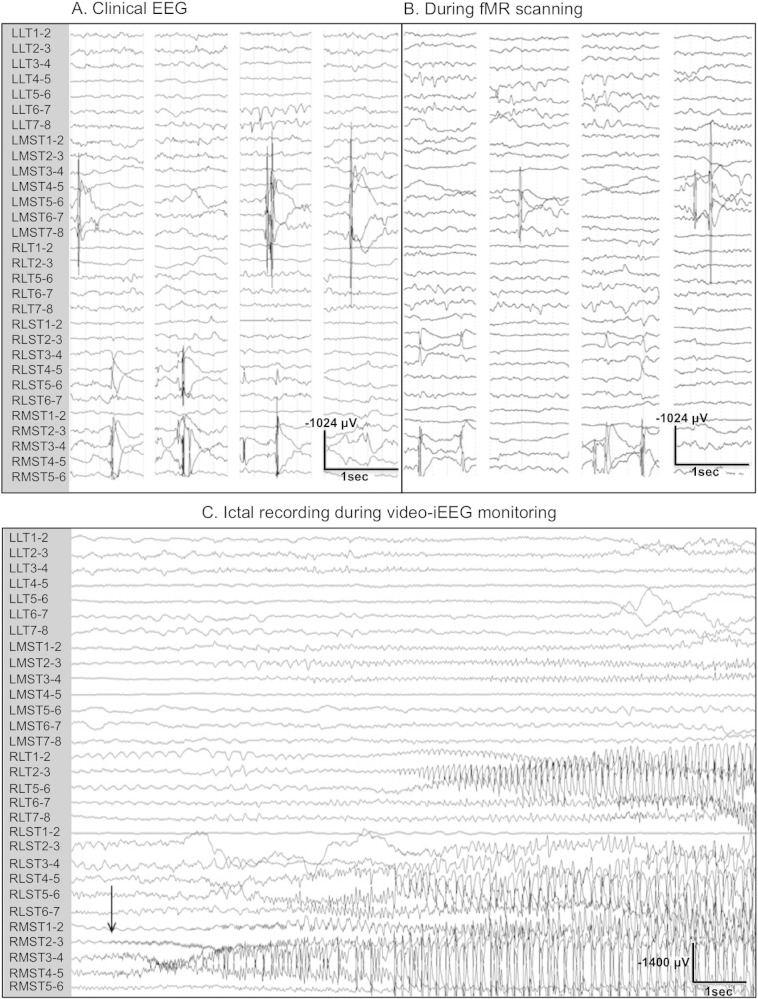
Objectives: Simultaneous scalp EEG-fMRI can identify hemodynamic changes associated with the generation of interictal epileptiform discharges (IEDs), and it has the potential of becoming a standard, non-invasive technique for pre-surgical assessment of patients with medically intractable epilepsy. This study was designed to assess the BOLD response to focal IEDs recorded via simultaneous intracranial EEG-functional MRI (iEEG-fMRI).
Methods: Twelve consecutive patients undergoing intracranial video EEG monitoring were recruited for iEEG-fMRI studies at 3 T. Depth, subdural strip, or grid electrodes were implanted according to our standard clinical protocol. Subjects underwent 10-60 min of continuous iEEG-fMRI scanning. IEDs were marked, and the most statistically significant clusters of BOLD signal were identified (Z-score 2.3, p value < 0.05). We assessed the concordance between the locations of the BOLD response and the IED. Concordance was defined as a distance <1.0 cm between the IED and BOLD response location. Negative BOLD responses were not studied in this project.
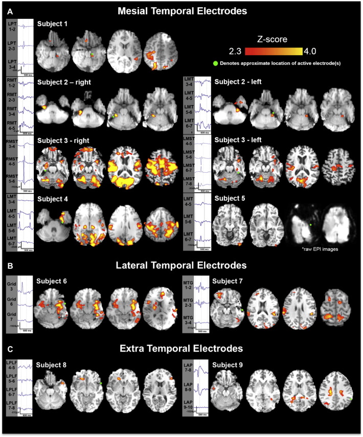
Results: Nine patients (7 females) with a mean age of 31 years (range 22-56) had 11 different types of IEDs during fMR scanning. The IEDs were divided based on the location of the active electrode contact into mesial temporal, lateral temporal, and extra-temporal. Seven (5 left) mesial temporal IED types were recorded in 5 patients (110-2092 IEDs per spike location). Six of these IEDs had concordant BOLD response in the ipsilateral mesial temporal structures, <1 cm from the most active contact. One of the two subjects with left lateral temporal IEDs had BOLD responses concordant with the location of the most active contact, as well other ipsilateral and contralateral sites. Notably, the remaining two subjects with extratemporal discharges showed no BOLD signal near the active electrode contact.
Conclusions: iEEG-fMRI is a feasible and low-risk method for assessment of hemodynamic changes of very focal IEDs that may not be recorded by scalp EEG. A high concordance rate between the location of the BOLD response and IEDs was seen for mesial temporal (6/7) IEDs. Significant BOLD activation was also seen in areas distant from the active electrode and these sites exhibited maximal BOLD activation in the majority of cases. This implies that iEEG-fMRI may further describe the areas involved in the generation of IEDs beyond the vicinity of the electrode(s).
Keywords: BOLD response; EEG-fMRI; Epileptiform discharge; IED, interictal epileptiform discharge; VEM, video-EEG monitoring.; intracranial EEG.
Figures
Similar articles
-
Mesial temporal lobe spiking reveals distinct patterns of blood oxygen level-dependent functional magnetic resonance imaging activation using simultaneous intracranial electroencephalography-functional magnetic resonance imaging.Epilepsia. 2024 Aug;65(8):2295-2307. doi: 10.1111/epi.18036. Epub 2024 Jun 7. Epilepsia. 2024. PMID: 38845414
-
Intracranial EEG-fMRI analysis of focal epileptiform discharges in humans.Epilepsia. 2012 Sep;53(9):1636-48. doi: 10.1111/j.1528-1167.2012.03601.x. Epub 2012 Aug 6. Epilepsia. 2012. PMID: 22881457
-
Localization of interictal discharge origin: A simultaneous intracranial electroencephalographic-functional magnetic resonance imaging study.Epilepsia. 2021 May;62(5):1105-1118. doi: 10.1111/epi.16887. Epub 2021 Mar 29. Epilepsia. 2021. PMID: 33782964
-
Role of specific interictal and ictal EEG onset patterns.Epilepsy Behav. 2025 Mar;164:110298. doi: 10.1016/j.yebeh.2025.110298. Epub 2025 Feb 7. Epilepsy Behav. 2025. PMID: 39922077 Review.
-
The impact of EEG/MEG signal processing and modeling in the diagnostic and management of epilepsy.IEEE Rev Biomed Eng. 2008;1:143-56. doi: 10.1109/RBME.2008.2008246. IEEE Rev Biomed Eng. 2008. PMID: 22274902 Review.
Cited by
-
Localizing Epileptic Foci Using Simultaneous EEG-fMRI Recording: Template Component Cross-Correlation.Front Neurol. 2021 Nov 15;12:695997. doi: 10.3389/fneur.2021.695997. eCollection 2021. Front Neurol. 2021. PMID: 34867704 Free PMC article.
-
Optimizing EEG Source Reconstruction with Concurrent fMRI-Derived Spatial Priors.Brain Topogr. 2022 May;35(3):282-301. doi: 10.1007/s10548-022-00891-3. Epub 2022 Feb 10. Brain Topogr. 2022. PMID: 35142957 Free PMC article.
-
Brain imaging in the assessment for epilepsy surgery.Lancet Neurol. 2016 Apr;15(4):420-33. doi: 10.1016/S1474-4422(15)00383-X. Epub 2016 Feb 24. Lancet Neurol. 2016. PMID: 26925532 Free PMC article. Review.
-
Tonic Resting State Hubness Supports High Gamma Activity Defined Verbal Memory Encoding Network in Epilepsy.Neuroscience. 2020 Jan 15;425:194-216. doi: 10.1016/j.neuroscience.2019.11.001. Epub 2019 Nov 28. Neuroscience. 2020. PMID: 31786346 Free PMC article.
-
Spike-related haemodynamic responses overlap with high frequency oscillations in patients with focal epilepsy.Brain. 2018 Mar 1;141(3):731-743. doi: 10.1093/brain/awx383. Brain. 2018. PMID: 29360943 Free PMC article.
References
Publication types
MeSH terms
Grants and funding
LinkOut - more resources
Full Text Sources
Other Literature Sources
Medical
