

Comparing a diffusion tensor and non-tensor approach to white matter fiber tractography in chronic stroke
- PMID: 25844329
- PMCID: PMC4375634
- DOI: 10.1016/j.nicl.2015.03.007
Comparing a diffusion tensor and non-tensor approach to white matter fiber tractography in chronic stroke
Abstract
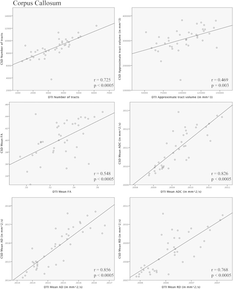
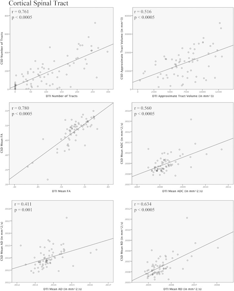
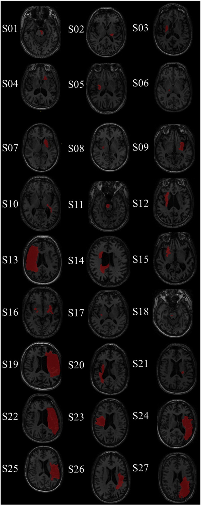
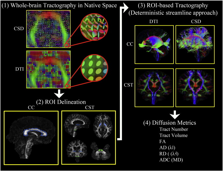
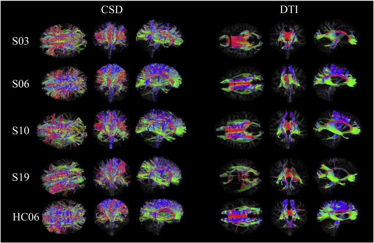
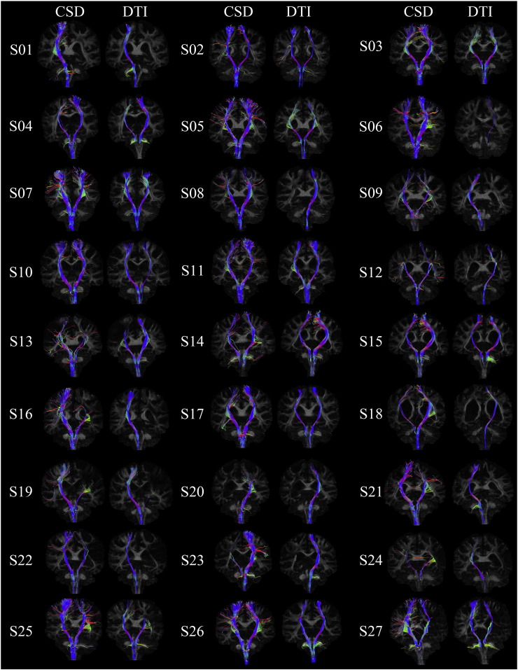
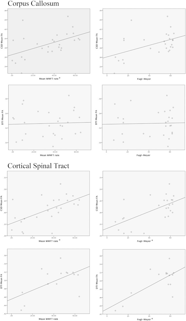
Diffusion tensor imaging (DTI)-based tractography has been used to demonstrate functionally relevant differences in white matter pathway status after stroke. However, it is now known that the tensor model is insensitive to the complex fiber architectures found in the vast majority of voxels in the human brain. The inability to resolve intra-voxel fiber orientations may have important implications for the utility of standard DTI-based tract reconstruction methods. Intra-voxel fiber orientations can now be identified using novel, tensor-free approaches. Constrained spherical deconvolution (CSD) is one approach to characterize intra-voxel diffusion behavior. In the current study, we performed DTI- and CSD-based tract reconstruction of the corticospinal tract (CST) and corpus callosum (CC) to test the hypothesis that characterization of complex fiber orientations may improve the robustness of fiber tract reconstruction and increase the sensitivity to identify functionally relevant white matter abnormalities in individuals with chronic stroke. Diffusion weighted magnetic resonance imaging was performed in 27 chronic post-stroke participants and 12 healthy controls. Transcallosal pathways and the CST bilaterally were reconstructed using DTI- and CSD-based tractography. Mean fractional anisotropy (FA), apparent diffusion coefficient (ADC), axial diffusivity (AD), and radial diffusivity (RD) were calculated across the tracts of interest. The total number and volume of reconstructed tracts was also determined. Diffusion measures were compared between groups (Stroke, Control) and methods (CSD, DTI). The relationship between post-stroke motor behavior and diffusion measures was evaluated. Overall, CSD methods identified more tracts than the DTI-based approach for both CC and CST pathways. Mean FA, ADC, and RD differed between DTI and CSD for CC-mediated tracts. In these tracts, we discovered a difference in FA for the CC between stroke and healthy control groups using CSD but not DTI. CSD identified ipsilesional CST pathways in 9 stroke participants who did not have tracts identified with DTI. Additionally, CSD differentiated between stroke ipsilesional and healthy control non-dominant CST for several measures (number of tracts, tract volume, FA, ADC, and RD) whereas DTI only detected group differences for number of tracts. In the stroke group, motor behavior correlated with fewer diffusion metrics derived from the DTI as compared to CSD-reconstructed ipsilesional CST and CC. CSD is superior to DTI-based tractography in detecting differences in diffusion characteristics between the nondominant healthy control and ipsilesional CST. CSD measures of microstructure tissue properties related to more motor outcomes than DTI measures did. Our results suggest the potential utility and functional relevance of characterizing complex fiber organization using tensor-free diffusion modeling approaches to investigate white matter pathways in the brain after stroke.
Keywords: Constrained spherical deconvolution; Diffusion tensor imaging; Diffusion weighted imaging; Motor outcome; Stroke.
Figures
References
Publication types
MeSH terms
Grants and funding
LinkOut - more resources
Full Text Sources
Other Literature Sources
Medical
Miscellaneous
