

Nanoscale strategies: treatment for peripheral vascular disease and critical limb ischemia
- PMID: 25844518
- PMCID: PMC5494973
- DOI: 10.1021/nn507269g
Nanoscale strategies: treatment for peripheral vascular disease and critical limb ischemia
Abstract
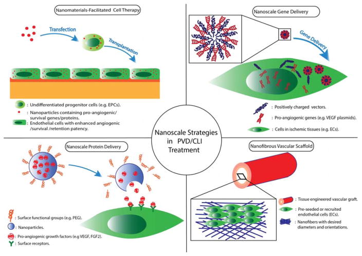
Peripheral vascular disease (PVD) is one of the most prevalent vascular diseases in the U.S. afflicting an estimated 8 million people. Obstruction of peripheral arteries leads to insufficient nutrients and oxygen supply to extremities, which, if not treated properly, can potentially give rise to a severe condition called critical limb ischemia (CLI). CLI is associated with extremely high morbidities and mortalities. Conventional treatments such as angioplasty, atherectomy, stent implantation and bypass surgery have achieved some success in treating localized macrovascular disease but are limited by their invasiveness. An emerging alternative is the use of growth factor (delivered as genes or proteins) and cell therapy for PVD treatment. By delivering growth factors or cells to the ischemic tissue, one can stimulate the regeneration of functional vasculature network locally, re-perfuse the ischemic tissue, and thus salvage the limb. Here we review recent advance in nanomaterials, and discuss how their application can improve and facilitate growth factor or cell therapies. Specifically, nanoparticles (NPs) can serve as drug carrier and target to ischemic tissues and achieve localized and sustained release of pro-angiogenic proteins. As nonviral vectors, NPs can greatly enhance the transfection of target cells with pro-angiogenic genes with relatively fewer safety concern. Further, NPs may also be used in combination with cell therapy to enhance cell retention, cell survival and secretion of angiogenic factors. Lastly, nano/micro fibrous vascular grafts can be engineered to better mimic the structure and composition of native vessels, and hopefully overcome many complications/limitations associated with conventional synthetic grafts.
Keywords: cell therapy; critical limb ischemia; gene delivery; growth factor; nanomaterials; nanomedicine; nanoparticles; peripheral vascular disease; stem cells; therapeutic angiogenesis; tissue engineering; vascular graft.
Conflict of interest statement
Figures
Similar articles
-
Allogeneic transplantation of programmable cells of monocytic origin (PCMO) improves angiogenesis and tissue recovery in critical limb ischemia (CLI): a translational approach.Stem Cell Res Ther. 2018 Apr 27;9(1):117. doi: 10.1186/s13287-018-0871-8. Stem Cell Res Ther. 2018. PMID: 29703251 Free PMC article.
-
Therapeutic angiogenesis in the management of critical limb ischemia: current concepts and review.Cardiol Rev. 2009 May-Jun;17(3):115-20. doi: 10.1097/CRD.0b013e318199e9b7. Cardiol Rev. 2009. PMID: 19384084 Review.
-
Therapeutic angiogenesis using zinc oxide nanoflowers for the treatment of hind limb ischemia in a rat model.Biomed Mater. 2021 Mar 26;16(4). doi: 10.1088/1748-605X/abebd1. Biomed Mater. 2021. PMID: 33657534
-
Concise Review: Cell Therapy for Critical Limb Ischemia: An Integrated Review of Preclinical and Clinical Studies.Stem Cells. 2018 Feb;36(2):161-171. doi: 10.1002/stem.2751. Epub 2018 Jan 3. Stem Cells. 2018. PMID: 29226477 Review.
-
Difference in Serum Endostatin Levels in Diabetic Patients with Critical Limb Ischemia Treated by Autologous Cell Therapy or Percutaneous Transluminal Angioplasty.Cell Transplant. 2018 Sep;27(9):1368-1374. doi: 10.1177/0963689718775628. Epub 2018 Jun 4. Cell Transplant. 2018. PMID: 29860903 Free PMC article.
Cited by
-
Bioengineering Cell Therapy for Treatment of Peripheral Artery Disease.Arterioscler Thromb Vasc Biol. 2024 Mar;44(3):e66-e81. doi: 10.1161/ATVBAHA.123.318126. Epub 2024 Jan 4. Arterioscler Thromb Vasc Biol. 2024. PMID: 38174560 Free PMC article. Review.
-
Accelerated Blood Clearance Phenomenon Reduces the Passive Targeting of PEGylated Nanoparticles in Peripheral Arterial Disease.ACS Appl Mater Interfaces. 2016 Jul 20;8(28):17955-63. doi: 10.1021/acsami.6b05840. Epub 2016 Jul 7. ACS Appl Mater Interfaces. 2016. PMID: 27340833 Free PMC article.
-
Stem cell-based drug delivery strategy for skin regeneration and wound healing: potential clinical applications.Inflamm Regen. 2023 Jun 30;43(1):33. doi: 10.1186/s41232-023-00287-1. Inflamm Regen. 2023. PMID: 37391780 Free PMC article. Review.
-
Therapeutic Biomaterial Approaches to Alleviate Chronic Limb Threatening Ischemia.Adv Sci (Weinh). 2021 Feb 8;8(7):2003119. doi: 10.1002/advs.202003119. eCollection 2021 Apr. Adv Sci (Weinh). 2021. PMID: 33854887 Free PMC article. Review.
-
Design and Application Strategies of Natural Polymer Biomaterials in Artificial Ovaries.Ann Biomed Eng. 2023 Mar;51(3):461-478. doi: 10.1007/s10439-022-03125-6. Epub 2023 Jan 11. Ann Biomed Eng. 2023. PMID: 36629950 Review.
References
-
- Ouriel K. Peripheral Arterial Disease. Lancet. 2001;358:1257–1264. - PubMed
-
- Minar E. Critical Limb Ischaemia. Hamostaseologie. 2009;29:102–9. - PubMed
-
- Norgren L, Hiatt WR, Dormandy JA, Nehler MR, Harris KA, Fowkes FG. Inter-Society Consensus for the Management of Peripheral Arterial Disease (Tasc II) J Vasc Surg. 2007;45(Suppl S):S5–67. - PubMed
Publication types
MeSH terms
Grants and funding
LinkOut - more resources
Full Text Sources
Other Literature Sources
Research Materials
Miscellaneous
