

Transcriptional profile of Mycobacterium tuberculosis replicating in type II alveolar epithelial cells
- PMID: 25844539
- PMCID: PMC4386821
- DOI: 10.1371/journal.pone.0123745
Transcriptional profile of Mycobacterium tuberculosis replicating in type II alveolar epithelial cells
Abstract
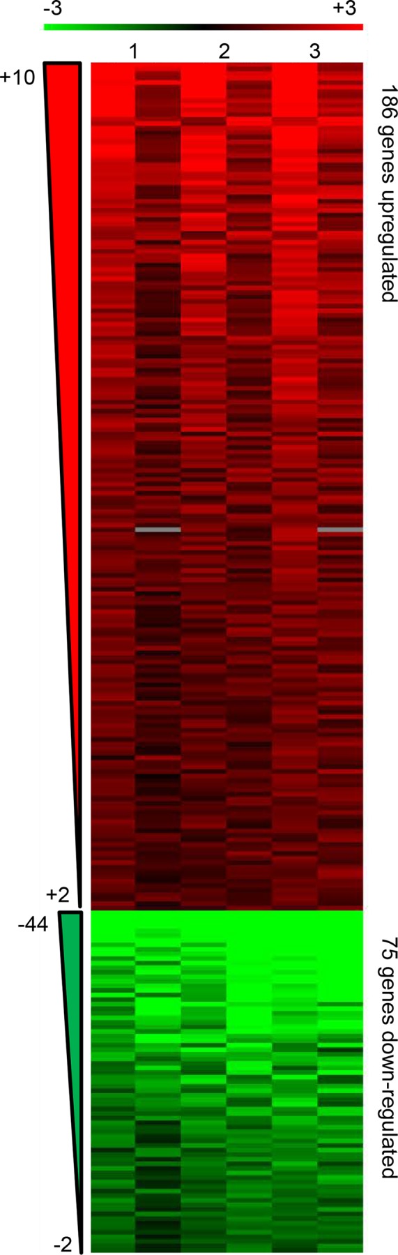
Mycobacterium tuberculosis (M. tb) infection is initiated by the few bacilli inhaled into the alveolus. Studies in lungs of aerosol-infected mice provided evidence for extensive replication of M. tb in non-migrating, non-antigen-presenting cells in the alveoli during the first 2-3 weeks post-infection. Alveoli are lined by type II and type I alveolar epithelial cells (AEC) which outnumber alveolar macrophages by several hundred-fold. M. tb DNA and viable M. tb have been demonstrated in AEC and other non-macrophage cells of the kidney, liver, and spleen in autopsied tissues from latently-infected subjects from TB-endemic regions indicating systemic bacterial dissemination during primary infection. M. tb have also been demonstrated to replicate rapidly in A549 cells (type II AEC line) and acquire increased invasiveness for endothelial cells. Together, these results suggest that AEC could provide an important niche for bacterial expansion and development of a phenotype that promotes dissemination during primary infection. In the current studies, we have compared the transcriptional profile of M. tb replicating intracellularly in A549 cells to that of M. tb replicating in laboratory broth, by microarray analysis. Genes significantly upregulated during intracellular residence were consistent with an active, replicative, metabolic, and aerobic state, as were genes for tryptophan synthesis and for increased virulence (ESAT-6, and ESAT-6-like genes, esxH, esxJ, esxK, esxP, and esxW). In contrast, significant downregulation of the DevR (DosR) regulon and several hypoxia-induced genes was observed. Stress response genes were either not differentially expressed or were downregulated with the exception of the heat shock response and those induced by low pH. The intra-type II AEC M. tb transcriptome strongly suggests that AEC could provide a safe haven in which M. tb can expand dramatically and disseminate from the lung prior to the elicitation of adaptive immune responses.
Conflict of interest statement
Figures
Similar articles
-
Transcriptional profiling of Mycobacterium tuberculosis replicating ex vivo in blood from HIV- and HIV+ subjects.PLoS One. 2014 Apr 22;9(4):e94939. doi: 10.1371/journal.pone.0094939. eCollection 2014. PLoS One. 2014. PMID: 24755630 Free PMC article.
-
Rv3351c, a Mycobacterium tuberculosis gene that affects bacterial growth and alveolar epithelial cell viability.Can J Microbiol. 2015 Dec;61(12):938-47. doi: 10.1139/cjm-2015-0528. Epub 2015 Oct 22. Can J Microbiol. 2015. PMID: 26492080
-
Mycobacterium tuberculosis infection causes different levels of apoptosis and necrosis in human macrophages and alveolar epithelial cells.Cell Microbiol. 2003 Sep;5(9):649-60. doi: 10.1046/j.1462-5822.2003.00312.x. Cell Microbiol. 2003. PMID: 12925134
-
Mycobacterium tuberculosis Primary Infection and Dissemination: A Critical Role for Alveolar Epithelial Cells.Front Cell Infect Microbiol. 2019 Aug 21;9:299. doi: 10.3389/fcimb.2019.00299. eCollection 2019. Front Cell Infect Microbiol. 2019. PMID: 31497538 Free PMC article. Review.
-
The mechanisms and consequences of the extra-pulmonary dissemination of Mycobacterium tuberculosis.Tuberculosis (Edinb). 2010 Nov;90(6):361-6. doi: 10.1016/j.tube.2010.08.005. Epub 2010 Sep 9. Tuberculosis (Edinb). 2010. PMID: 20829117 Review.
Cited by
-
In Vitro Models for Studying Respiratory Host-Pathogen Interactions.Adv Biol (Weinh). 2021 Jun;5(6):e2000624. doi: 10.1002/adbi.202000624. Epub 2021 May 4. Adv Biol (Weinh). 2021. PMID: 33943040 Free PMC article. Review.
-
Transcriptome analysis of mycobacteria in sputum samples of pulmonary tuberculosis patients.PLoS One. 2017 Mar 10;12(3):e0173508. doi: 10.1371/journal.pone.0173508. eCollection 2017. PLoS One. 2017. PMID: 28282458 Free PMC article.
-
miR-296-5p Inhibits the Secretion of Pulmonary Surfactants in Pulmonary Epithelial Cells via the Downregulation of Wnt7b/β-Catenin Signaling.Biomed Res Int. 2021 Jan 5;2021:4051504. doi: 10.1155/2021/4051504. eCollection 2021. Biomed Res Int. 2021. PMID: 33490270 Free PMC article.
-
Inflammation, infection and depression: an evolutionary perspective.Evol Hum Sci. 2019 Dec 9;1:e14. doi: 10.1017/ehs.2019.15. eCollection 2019. Evol Hum Sci. 2019. PMID: 37588396 Free PMC article.
-
AmpliSeq transcriptome analysis of human alveolar and monocyte-derived macrophages over time in response to Mycobacterium tuberculosis infection.PLoS One. 2018 May 30;13(5):e0198221. doi: 10.1371/journal.pone.0198221. eCollection 2018. PLoS One. 2018. PMID: 29847580 Free PMC article.
References
-
- Hestvik AL, Hmama Z, Av-Gay Y. Mycobacterial manipulation of the host cell. FEMS Microbiol Rev. 2005. November;29(5):1041–50. - PubMed
-
- Crandall ED, Kim KJ. Alveolar epithelial barrier properties In: Crystal RJ, West JB, editors. The Lung: Scientific Foundations. New York, NY: Raven Press; 1991.
-
- Crystal RJ. Alveolar Macrophages In: Crystal RJ, West JB, editors. The Lung: Scientific Foundations. New York, NY: Raven Press; 1991. p. 527–38.
Publication types
MeSH terms
Substances
Associated data
- Actions
Grants and funding
LinkOut - more resources
Full Text Sources
Other Literature Sources
Molecular Biology Databases
