. 2015 Apr 30;43(8):4087-97.
doi: 10.1093/nar/gkv301. Epub 2015 Apr 6.

Molecular crowding enhances facilitated diffusion of two human DNA glycosylases

Affiliations

Molecular crowding enhances facilitated diffusion of two human DNA glycosylases

Shannen L Cravens et al. Nucleic Acids Res. .

Abstract

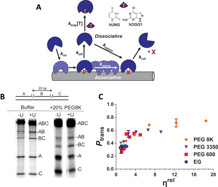
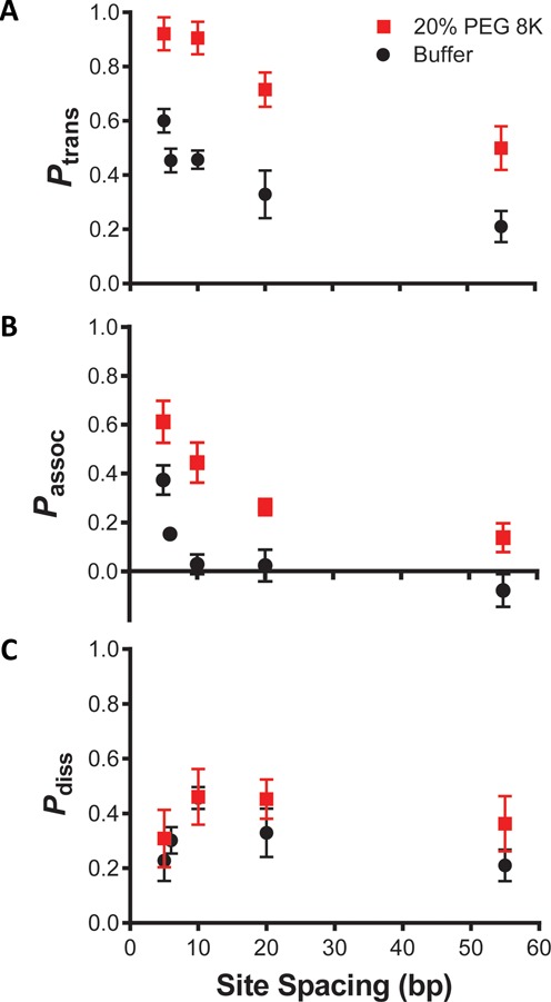
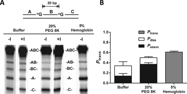
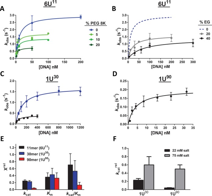
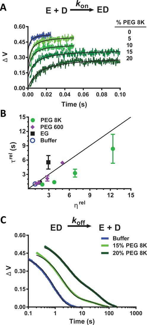
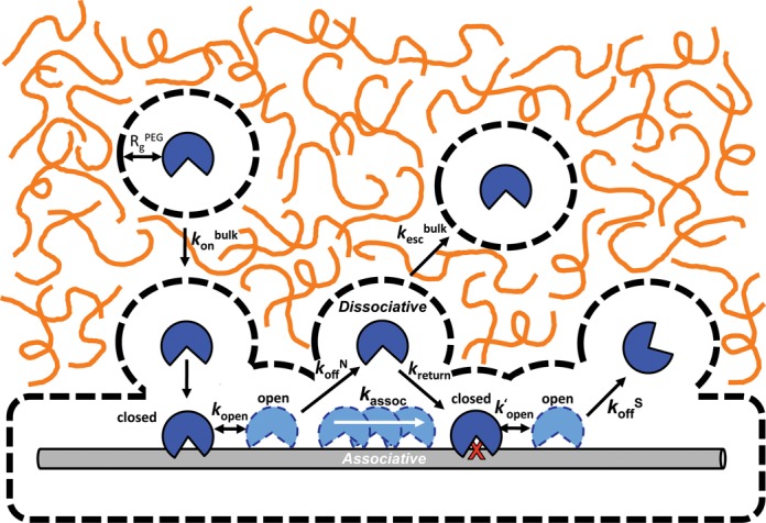
Intracellular space is at a premium due to the high concentrations of biomolecules and is expected to have a fundamental effect on how large macromolecules move in the cell. Here, we report that crowded solutions promote intramolecular DNA translocation by two human DNA repair glycosylases. The crowding effect increases both the efficiency and average distance of DNA chain translocation by hindering escape of the enzymes to bulk solution. The increased contact time with the DNA chain provides for redundant damage patrolling within individual DNA chains at the expense of slowing the overall rate of damaged base removal from a population of molecules. The significant biological implication is that a crowded cellular environment could influence the mechanism of damage recognition as much as any property of the enzyme or DNA.


Figures

Figure 1.
Figure 1.
Approach for measuring site transfer probabilities of hUNG and hOGG1. (a) The multistep search and repair pathway used by DNA glycosylases. The pathway begins with association to form a nonspecific DNA complex followed by associative and dissociative transfer steps along the DNA chain until a damaged site is located. Site transfer probability measurements involve the use of substrates with two sites of known separation and the determination of the likelihood that an enzyme molecule that produces product (P) at one site successfully transfers to the second site (X) without dissociating to bulk solution (3,30,31). The method has been modified to include a ‘molecular clock’ where a small molecule trap [T = U for hUNG, or I for hOGG1] is used to prevent transfers by the dissociative pathway, allowing for direct measurement of associative transfers only (see text). (b) Phosphorimages of the products derived from reaction of hUNG with a 90mer substrate (S20U) with two uracils spaced 20bp apart in dilute buffer and in buffer containing 20% PEG 8K (±U denotes the presence and absence of uracil trap). The increased transfers in the presence of 20% PEG 8K are indicated by the increased levels of the (A) and (C) bands (double excision fragments) as compared to AB and BC bands (single excision fragments). (c) The transfer probabilities between uracil sites spaced 20 bp apart (measured at 37°C) as a function of relative viscosity (ηrel = ηcrowder/ηbuffer) for a series of PEG polymers. Relative viscosities of these cosolutes were obtained from reference (29). The Ptrans value for buffer alone is indicated by the black open circle.
Figure 2.
Figure 2.
Effect of 20% PEG 8K on the intramolecular site transfer probability between uracil sites of variable spacing. All data obtained from dilute buffer conditions (black) have been previously reported and are displayed here for comparison (30). (a) Overall site transfer probabilities (Ptrans) as a function of spacing length between uracil sites in the presence (red) and absence (black) of PEG 8K. (b) The increased contribution of the associative transfer pathway (Passoc) and the longer associative transfer distances in the presence of PEG 8K were determined using high concentrations of uracil trap (10 and 20 mM). (c) A smaller increase in the probability of dissociative transfers (Pdiss = PtransPassoc) was observed for all site spacings in the presence of PEG 8K.
Figure 3.
Figure 3.
Effect of 20% PEG 8K and 5% hemoglobin on the site transfer probability of hOGG1 over a 20 bp spacing (Ptrans). (a) Phosphorimages of polyacrylamide gels showing the separation of the reaction products generated from excision of 8-oxoG. The presence or absence of the molecular trap (Figure 1) is indicated by ±I. (b) Partitioning of the total transfers between the associative (Passoc) and dissociative (Pdiss) pathways. Error bars show the SD of three replicate determinations of Ptrans. Overall Ptrans was found to increase when PEG 8K was added, primarily due to an increase in the relative contribution of the associative pathway. A similar increase in Ptrans was observed upon addition of hemoglobin, though the contributions of Passoc and Pdiss could not be discerned due to precipitation upon addition of the trap.
Figure 4.
Figure 4.
The effect of molecular crowding agents on the steady-state kinetics of hUNG acting on short (11 and 30 bp) and long (90 bp) duplex DNA substrates. The kinetic parameters obtained from all of the data sets in this figure are reported in Supplementary Table S5. (a) Effect of 5–20% PEG 8K on the initial rates of reaction of hUNG with a DNA hairpin substrate containing an 11 bp stem (6U11). (b) Effect of 20–40% EG on the initial reaction rates of hUNG with 6U11. For comparison, the dashed blue line is the curve for dilute buffer shown in panel (a). (c) Effect of 20% PEG 8K (black) on the initial reaction rates of hUNG with a 30 mer duplex (1U30) in comparison to dilute buffer (blue). (d) Initial rates of uracil excision from the 90mer duplex (1U90) in the presence of 20% PEG 8K. Previously published data with the same substrate in the absence of PEG 8K are reported in Supplementary Table S5. (e) The relative effect of 20% PEG 8K on the steady-state kinetic parameters (Xrel) for hUNG acting on short (6U11 and 1U30) and long (1U90) duplex DNA substrates. Xrel is defined as the kinetic parameter obtained in the presence of PEG 8K divided by that obtained in buffer alone (see Supplementary Table S5). (f) The salt-dependent change in kcat for 30 bp (1U30) and 90 bp (1U90) DNA substrates in the presence of molecular crowding agents. The effect of 20% PEG 8K on kcat for both substrates was determined in presence of high salt (75 mM total cation concentration, gray bars). For comparison, the black bars show the kcat values determined at low salt (22 mM cation concentration, see Supplementary Figure S5). The parameter kcatrel is defined as the kcat value determined in the presence of 20% PEG 8K divided by the value obtained using buffer alone. The kcat values for both 1U30 and 1U90 dropped ∼2-fold upon addition of 20% PEG 8K (1U30): kcat decreased from 2.1 ± 0.9 to 1.2 ± 0.5 s−1; 1U90kcat decreased from 10.5 ± 0.4 to 5 ± 2 s−1. The similar effect of PEG 8K for both substrates at high, but not low salt, supports the proposal that rate-limiting associative transfers limit turnover of the large substrate at low salt. The differences in the absolute kcat values for each substrate are due to sequence dependent differences in the steady-state turnover rate (5,49).
Figure 5.
Figure 5.
Influence of PEG 8K on association kinetics of hUNG from specific DNA (DS) determined by stopped-flow 2-aminopurine fluorescence measurements at 20°C. (a) Kinetic traces for the second-order association of hUNG (200 nM) with DS (200 nM) in dilute buffer (blue) and in the presence of 5–20% PEG 8K. Traces are displaced along the y-axis for clarity. The second-order rate constants (kon) are reported in Supplementary Table S5. (b) Effect of relative viscosity (ηrel = ηcrowder/ηbuffer) on the relative association times (τrel = τcrowder/τbuffer, where τ = 1/kon) for hUNG and specific DNA (DS). The data points correspond to dilute buffer (blue) and increasing concentrations of EG (black), PEG 600 (purple) and PEG 8K (green). Relative viscosities of these cosolutes were obtained from (29). The theoretical line shows the expected dependence of the association times based solely on the increases in the relative viscosity of the solutions as expected from Stokes–Einstein behavior. (c) Effect of PEG 8K on the dissociation rate of hUNG from a specific site (DS). Kinetic traces are shown in a semi-log format. The dissociation kinetics follow a single exponential in buffer alone (blue), and a double exponential in the presence of 15% (light green) and 20% PEG 8K (dark green). The second slower exponential that appears in the presence of PEG 8K is deemed a fluorescence artifact as detailed in the Supplemental Methods. The kinetic parameters are reported in Supplementary Table S6.
Figure 6.
Figure 6.
General schematic of how the introduction of molecular crowders (orange lines) can influence individual steps in the DNA glycosylase damage search pathway. These steps including the rate of diffusion to the DNA chain (kon), the lifetime of nonspecific (1/koffN) and specific (1/koffS) DNA complexes, the probability of associative and dissociative transfers between damage sites, and changes in the rate of product release (rate-limiting for kcat). Dashed lines represent the sizes of the depletion layers surrounding the protein and DNA where the PEG 8K polymer, but not solvent, is excluded. For large DNA molecules, association is limited by translation of the protein through the crowded solution (konbulk) until their depletion layers overlap and association proceeds within a low viscosity environment. Also depicted are highly dynamic closed-to-open conformational changes in hUNG and hOGG1 that accompany nonspecific DNA binding (42,50); only the open state is viewed competent for translocation (50). Release of each enzyme from the product requires an even larger closed-to-open transition that has been shown to be at least partly rate-limiting for turnover of hUNG (k′open) (46). Image is drawn to scale using a DNA duplex of 2 nm as a scale reference. The depletion layer size in the dilute regime for PEG 8K is shown (equivalent to RgPEG, see Supplemental Information).

References

    1. Blainey P.C., Oijen A.M.V., Banerjee A., Verdine G.L., Xie X.S. A base-excision DNA-repair protein finds intrahelical lesion bases by fast sliding in contact with DNA. Proc. Natl. Acad. Sci. U.S.A. 2006;103:5752–5757. - PMC - PubMed
    1. Blainey P.C., Luo G., Kou S.C., Mangel W.F., Verdine G.L., Bagchi B., Xie X.S. Nonspecifically bound proteins spin while diffusing along DNA. Nat. Struct. Mol. Biol. 2009;16:1224–1229. - PMC - PubMed
    1. Porecha R.H., Stivers J.T. Uracil DNA glycosylase uses DNA hopping and short-range sliding to trap extrahelical uracils. Proc. Natl. Acad. Sci. U.S.A. 2008;105:10791–10796. - PMC - PubMed
    1. Hedglin M., O'Brien P.J. Human alkyladenine DNA glycosylase employs a processive search for DNA damage. Biochemistry. 2008;47:11434–11445. - PMC - PubMed
    1. Krosky D.J., Song F., Stivers J.T. The origins of high-affinity enzyme binding to an extrahelical DNA base. Biochemistry. 2005;44:5949–5959. - PubMed

Publication types

LinkOut - more resources