. 2015 Oct 1;523(14):2138-60.
doi: 10.1002/cne.23783. Epub 2015 Jun 2.

Spinal cord neuron inputs to the cuneate nucleus that partially survive dorsal column lesions: A pathway that could contribute to recovery after spinal cord injury

Affiliations

Spinal cord neuron inputs to the cuneate nucleus that partially survive dorsal column lesions: A pathway that could contribute to recovery after spinal cord injury

Chia-Chi Liao et al. J Comp Neurol. .

Abstract

Dorsal column lesions at a high cervical level deprive the cuneate nucleus and much of the somatosensory system of its major cutaneous inputs. Over weeks of recovery, much of the hand representations in the contralateral cortex are reactivated. One possibility for such cortical reactivation by hand afferents is that preserved second-order spinal cord neurons reach the cuneate nucleus through pathways that circumvent the dorsal column lesions, contributing to cortical reactivation in an increasingly effective manner over time. To evaluate this possibility, we first injected anatomical tracers into the cuneate nucleus and plotted the distributions of labeled spinal cord neurons and fibers in control monkeys. Large numbers of neurons in the dorsal horn of the cervical spinal cord were labeled, especially ipsilaterally in lamina IV. Labeled fibers were distributed in the cuneate fasciculus and lateral funiculus. In three other squirrel monkeys, unilateral dorsal column lesions were placed at the cervical segment 4 level and tracers were injected into the ipsilateral cuneate nucleus. Two weeks later, a largely unresponsive hand representation in contralateral somatosensory cortex confirmed the effectiveness of the dorsal column lesion. However, tracer injections in the cuneate nucleus labeled only about 5% of the normal number of dorsal horn neurons, mainly in lamina IV, below the level of lesions. Our results revealed a small second-order pathway to the cuneate nucleus that survives high cervical dorsal column lesions by traveling in the lateral funiculus. This could be important for cortical reactivation by hand afferents, and recovery of hand use.

Keywords: cortical reactivation; cuneate nucleus; dorsal column lesion; lateral funiculus; second-order spinal cord pathway.


Conflict of interest statement

Conflict of interest statement: The authors have no conflict of interest.

Figures

Figure 1
Figure 1
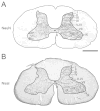
Transverse sections of the cervical spinal cord (C5) showing the laminae I–X of grey matter with (A) NeuN or (B) Nissl staining in squirrel monkeys. Laminae follow those portrayed in Sengal et al. (2012). Scale bar is 1 mm.
Figure 2
Figure 2
Distributions of spinal cord neurons labeled by injections of cholera toxin subunit B (CTB) into the dorsal column nuclei of owl monkey 11–18. A. CTB was injected into the cuneate nucleus on the left side and the gracile nucleus on the right side. The injection site in the left brainstem also involves the cuneate fasciculus and that in the right side involves the gracile nucleus with some lateral spread into the cuneate nucleus and fasciculus. Yellow and blue lines depict cores and halos of CTB injections. B. A large number of CTB-labeled neurons were found in the dorsal horn at a high cervical level C3 (box in D). The labeled neurons are marked by white circles. C. A large number of CTB-labeled neurons were found in the dorsal horn at a low cervical level C6 (box in D). D. A great number of CTB-labeled neurons (solid black circles) are primarily distributed in the dorsal horn of cervical spinal cord ipsilateral to the cuneate nucleus injection. A smaller number of CTB-labeled neurons appear on the right side of the cervical spinal cord. E. The percentages of the CTB-labeled neurons in laminae I–X across cervical segments C1–C8 ipsilateral to the injection site suggest that the labeled neurons are extensively distributed throughout the cervical spinal cord (red). Within each cervical segment, the CTB-labeled neurons are primarily located in the lamina IV and small numbers of labeled neurons are distributed in laminae V–VI and III (blue; shown as percentages in each segment). “n” represents the total number identified in each segment. Scale bar is 1 mm in A and 100 μm in BC. Cu, cuneate nucleus; Gr, gracile nucleus.
Figure 3
Figure 3
Distributions of spinal cord neurons labeled by injections of CTB into the cuneate nucleus of squirrel monkey 11–30. A. The CTB injection site mainly involves the cuneate nucleus and also the overlying cuneate fasciculus of the left brainstem. B. A large number of CTB-labeled neurons are located at a high cervical level C3 (box in D). C. A comparable number of CTB-labeled neurons are distributed at a low level C6 (box in D). D. The CTB-labeled neurons are mainly located in the medial aspect of the dorsal horn of the cervical spinal cord ipsilateral to the injection site, with a small number of CTB-labeled neurons on the contralateral side. E. The percentages of the CTB-labeled neurons in laminae I–X across cervical segments C1–C8 ipsilateral to the injection site, suggesting labeled neurons were extensively distributed throughout the cervical spinal cord. The CTB-labeled neurons are mainly located in the lamina IV and to a lesser percentage in the lamina III in each segment. Scale bar is 0.5 mm in A and 100 μm in BC.
Figure 4
Figure 4
Distributions of spinal cord neurons labeled by injections of CTB into the cuneate nucleus of squirrel monkey 12-04. A. The CTB injection site mainly involves the cuneate nucleus and overlying cuneate fasciculus of the left brainstem. B. A large number of CTB-labeled neurons are located at a high cervical level C3 (box in D). C. A comparable number of CTB-labeled neurons are distributed at a low level C6 (box in D). D. The CTB-labeled neurons are mainly located in the dorsal horn of the cervical spinal cord ipsilateral to the injection site, with a small number of CTB-labeled neurons on the contralateral side. E. The percentages of the CTB-labeled neurons in laminae I–X across cervical segments C1–C8 ipsilateral to the injection site. A great percentage of CTB-labeled neurons are located in the C1 and slightly lesser percentages of CTB-labeled neurons are distributed in C2 to C8. Within each segment, the majority of CTB-labeled neurons are located in the lamina IV and to a lesser percentage in the lamina III. Scale bar is 0.5 mm in A and 100 μm in BC.
Figure 5
Figure 5
Locations of axons connecting the cuneate nucleus and cervical spinal cord labeled by injection of fluoro-ruby (FR) into the cuneate nucleus of squirrel monkey 11–30. A. The FR injection site is confined to the cuneate nucleus. B. A great number of FR-labeled fibers are located across the depth of the cuneate fasciculus (box in D). The labeled-fibers are marked by arrowheads. C. A small number of FR-labeled fibers are located in the dorsal portion of the lateral funiculus (box in D). D. The distributions of FR-labeled fibers (red) are mainly in the dorsal column and fewer of them are in the lateral funiculus. The densities of labeled fibers are reduced toward caudal segments from C2 to C5. The FR-labeled neurons are mainly distributed in the dorsal horn of the cervical spinal cord ipsilateral to the injection site, and few of the labeled neurons are located in the contralateral side. Scale bar is 0.5 mm in A and 250 μm in BC.
Figure 6
Figure 6
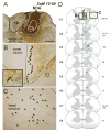
Locations of axons connecting the cuneate nucleus and cervical spinal cord labeled by injection of biotinylated dextran amine (BDA) into the cuneate nucleus of squirrel monkey 12-04. A. The BDA injection site is confined to the cuneate nucleus and the dense uptake zone extends slightly beyond the ventral border of cuneate nucleus. B. A large number of BDA-labeled fibers are located across the depth of the cuneate fasciculus (box in D). The labeled fibers are marked by arrowheads. The BDA injection labeled cells (arrow in the inset) and axon terminals (black circle in the inset) in the ipsilateral cervical spinal cord. C. A small number of BDA-labeled fibers (green) are located dorsally in the lateral funiculus (box in D). D. The distributions of BDA-labeled fibers are mainly in the cuneate fasciculus and fewer of them are in the lateral funiculus. The densities of labeled fibers are reduced toward caudal segments from C1 to C5. Only a few BDA-labeled neurons are located in the dorsal horn of the cervical spinal cord ipsilateral to the injection site. Scale bar is 0.5 mm in A, 250 μm in BC and 50 μm in the inset of B.
Figure 7
Figure 7
A complete dorsal column lesion deactivated the contralateral somatosensory cortex of squirrel monkey 12–52. A diagram showing the ascending pathways from the dorsal column and lateral funiculus of spinal cord to the cuneate nucleus in the brainstem, the ventroposterior nucleus (VP) in the thalamus, and up to the primary somatosensory area 3b. Two weeks after a complete dorsal column lesion at the C4 level (shown in black shading), multiunit recordings revealed that neurons throughout the contralateral hand representation of area 3b failed to respond to touches on the hand (penetrations marked with ×). However, neurons in the face representation of area 3b remained responsive. CS, central sulcus; GR, good response; M, medial; NR, no response; R, rostral; WR, weak response; vWR, very weak response. DCL, dorsal column lesion. Scale bar is 1 mm.
Figure 8
Figure 8
Distributions of spinal cord neurons labeled by injections of CTB into the cuneate nucleus after a complete dorsal column lesion at C4 of squirrel monkey 12–52. A. The CTB injection core site mainly involves the cuneate nucleus with a slight spread beyond the ventral border of cuneate nucleus. B. A large number of CTB-labeled neurons are located at a high cervical level C2 (box in D). C. A small number of CTB-labeled neurons are located at a low level C5 (box in D). D. Most of the CTB-labeled neurons are located in the dorsal horn of cervical spinal cord C1–C3. Below the dorsal column lesion level, a small numbers of CTB-labeled neurons are scattered in the cervical segments C4–C8. Few CTB-labeled neurons are distributed in the contralateral side. E. The percentages of the CTB-labeled neurons in laminae I–X across cervical segments C1–C8 ipsilateral to the injection site. Large percentages of CTB-labeled neurons are in C1 to C3, and significantly reduced percentages of labeled neurons are in C4 to C8 (p = 0.005, t-test). The CTB-labeled neurons are primarily distributed in the lamina IV, with a lesser amount in lamina V–VI above the lesion level. Below the lesion level, the CTB-labeled neurons tended to locate in the lamina IV but also scattered in other spinal laminae. Scale bar is 0.5 mm in A and 100 μm in BC.
Figure 9
Figure 9
A nearly complete dorsal column lesion deactivated the contralateral somatosensory cortex of squirrel monkey 13–17. A. The lesion site is at C4 in the spinal cord involving most of the right dorsal column, the medioventral region of the left dorsal column, medial regions of the dorsal horn on both sides, and the central canal region. B. Two weeks after the lesion, multiunit recording from microelectrode penetrations showing that the majority of neurons in the penetrations in the hand representation were unresponsive to touch. Neurons in 4 penetrations responded to peripheral stimuli with weak responses. The microelectrode penetrations in the face representation detected good or weak responses. The yellow, meshed shading marks a penetration with neurons responsive to hairs on dorsal digit 1, and the orange, meshed shadings mark locations with neurons responsive to hairs on radial hand. Scale bar is 1 mm in B. Please see Figure 7 for other conventions.
Figure 10
Figure 10
Distributions of spinal cord neurons labeled by an injection of CTB into the cuneate nucleus after a complete dorsal column lesion at C4 of squirrel monkey 13–17. A. The CTB injection core site mainly involves the cuneate nucleus and overlying cuneate fasciculus with a slight spread into the lateral aspect of the gracile nucleus. B. A large number of CTB-labeled neurons are located at a high cervical level C3 (box in D). C. A small number of CTB-labeled neurons are located at a low level C6 (box in D). D. The majority of CTB-labeled neurons are located in the dorsal horn of cervical spinal cord C1–C3. Below the dorsal column lesion level, a small number of CTB-labeled neurons are scattered in the cervical segments C4–C8. Few CTB-labeled neurons are distributed in the contralateral side. E. The percentages of the CTB-labeled neurons in laminae I–X across cervical segments C1–C8 ipsilateral to the injection site. Most CTB-labeled neurons are in C1 to C3. Significantly reduced percentages of labeled neurons are observed in C4 to C8 (p = 0.002, t-test). The CTB-labeled neurons are primarily distributed in the lamina IV, to a lesser amount in the lamina V–VI above the lesion level. Below the lesion level, the CTB-labeled neurons are mainly located in the lamina IV but are also in other spinal laminae. Scale bar is 0.5 mm in A and 100 μm in BC.
Figure 11
Figure 11
Terminations of peripheral afferents in the dorsal horn of spinal cord and cuneate nucleus in the brainstem after an incomplete lesion of the right cuneate fasciculus in squirrel monkey 13-01. Terminations of primary afferents in the cuneate nucleus were revealed by choleratoxin subunit B conjugated with wheat germ agglutinin-horseradish peroxide (B-HRP) injections into distal digits 1, 3 and 5 of both hands. A. The B-HRP-labeled axonal terminals are located in the dorsal horn of C5, C6 and C7 on the two sides (A1, A2 and A3; white arrowheads), corresponding to the peripheral inputs from digits 1, 3 and 5. The lesion site at C4 of the right side was restricted to the area between the posterior median sulcus and the posterior intermediate sulcus to the bottom of the dorsal column, which mainly involved the cuneate fasciculus. B. Photomicrographs showing the B-HRP-labeled axonal terminals (white arrowheads) form patches in the cuneate nucleus of the left brainstem throughout rostrocaudal sections. Small foci of B-HRP labeled terminals are scattered in the cuneate nucleus on the right side. C. Bar graphs showing the total surface area of B-HRP labeled foci at caudal to rostral levels relative to the obex in the cuneate nucleus on the left (black) and right (gray) sides. The values of distance are measured in millimeters caudally (negative numbers) in reference to the obex. D. The B-HRP labeled area on the lesioned side (0.01 mm2) of the cuneate nucleus is significantly less than the labeled area on the control side (0.11 mm2; p < 0.001, t-test), indicating an estimated completeness of 91% of dorsal column lesion (consistent with the estimate from transverse sections through the lesion site). Scale bar is 1 mm in A1A3 and is 1 mm in B.
Figure 12
Figure 12
Microelectrode multiunit recordings in the area 3b and adjoining areas 3a and 1 contralateral to the dorsal column lesion of squirrel monkey 13-01. A. In the hand representations, most electrode penetrations had neurons with weak or very weak responses to peripheral stimuli although few of them had neurons with good responses. A few penetrations had unresponsive neurons. The representations of digits 1–5 (outlined colored regions) were arranged in a normal mediolateral arrangement. The palm representation was located caudal to the digit representations. Penetrations within territories marked by meshed shading had neurons with receptive fields on the dorsal hairy hand. Others were on the glabrous hand. All penetrations in the face representation had neurons with very good or good responses. B. Examples of shifts of receptive fields along the mediolateral (letters) and rostrocaudal (numbers) rows of recording sites (red & blue). Scale bar is 1 mm in A. Please see Figure 7 for other conventions.
Figure 13
Figure 13
Distributions of spinal cord neurons labeled by injections of CTB into the cuneate nucleus after a complete dorsal column lesion at C4 of squirrel monkey 13-01. A. The CTB injection core site mainly involves the cuneate nucleus and overlying cuneate fasciculus. The dense uptake zone slightly spreads beyond the ventral border of cuneate nucleus. B. A great number of CTB-labeled neurons are located at a high cervical level C2 (box in D). C. A small number of CTB-labeled neurons are located below the lesion level C4 (box in D). D. The majority of CTB-labeled neurons are concentrated in the dorsal horn of cervical spinal cord C1–C3. A small number of CTB-labeled neurons are scattered in the cervical segments C4–C8 below the level of lesion. A few CTB-labeled neurons are distributed on the contralateral side. E. The percentages of the CTB-labeled neurons in laminae I–X across cervical segments C1–C8 ipsilateral to the injection site. Large percentages of CTB-labeled neurons are in C1 to C3. Significantly reduced percentages of labeled neurons are observed in C4 to C8 (p < 0.001, t-test). The CTB-labeled neurons are primarily distributed in the lamina IV, and to a lesser amount in lamina V–VI and III above the lesion level. Below the lesion level, the CTB-labeled neurons are mainly located in lamina V–VI and IV but are also scattered in other spinal laminae. Scale bar is 0.5 mm in A and 100 μm in BC.
Figure 14
Figure 14
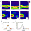
Summary of locations of spinal cord neurons labeled by the injections of CTB into the dorsal column nuclei in normal (n = 3) and dorsal column lesioned monkeys (n = 3). A. 2D heatmaps showing that the majority of CTB-labeled neurons are located in the lamina IV throughout the cervical spinal cord in normal monkeys. In the three dorsal column lesioned monkeys, the CTB-labeled neurons are concentrated in the lamina IV of C1 to C3. Small percentages of CTB-labeled neurons remain below the level of lesion C4 to C8. B. Comparisons of the percentages of labeled neurons in laminae I–X of C1–C3 above the lesion between the control and dorsal column lesion cases. The dorsal column lesion did not alter the laminar distributions (p = 0.309, Two Way Anova). C. Comparisons of the percentages of labeled neurons in laminae I–X of C4–C8 that are below the lesion between the two groups. The dorsal column lesions altered the overall distributions in laminae I–X (p < 0.001, Two Way Anova with post hoc Student-Newman-Keul Method). Although the percentages of labeled neurons remain high in the lamina IV, the percentages of labeled neurons in laminae IV is significantly reduced below the lesion. This allows the small number of neurons in lamina V–VI and VII to be proportionately significantly greater. Data in B and C are presented by mean ± SEM. * p < 0.05.

Similar articles

Cited by

References

    1. Balaram P, Hackett TA, Kaas JH. Differential expression of vesicular glutamate transporters 1 and 2 may identify distinct modes of glutamatergic transmission in the macaque visual system. J Chem Neuroanat. 2013;50–51:21–38. - PMC - PubMed
    1. Balaram P, Kaas JH. Towards a unified scheme of cortical lamination for primary visual cortex across primates: insights from NeuN and VGLUT2 immunoreactivity. Front Neuroanat. 2014;8:81. - PMC - PubMed
    1. Baldwin MK, Balaram P, Kaas JH. Projections of the superior colliculus to the pulvinar in prosimian galagos (Otolemur garnettii) and VGLUT2 staining of the visual pulvinar. J Comp Neurol. 2013;521(7):1664–1682. - PMC - PubMed
    1. Bennett GJ, Nishikawa N, Lu GW, Hoffert MJ, Dubner R. The morphology of dorsal column postsynaptic spinomedullary neurons in the cat. J Comp Neurol. 1984;224(4):568–578. - PubMed
    1. Bowes C, Burish M, Cerkevich C, Kaas J. Patterns of cortical reorganization in the adult marmoset after a cervical spinal cord injury. J Comp Neurol. 2013;521(15):3451–3463. - PMC - PubMed

Publication types

MeSH terms