Review
. 2015 Sep;45(10):1431-9.
doi: 10.1007/s00247-015-3336-6. Epub 2015 Apr 7.

Multidetector computed tomography pulmonary angiography in childhood acute pulmonary embolism

Affiliations
Review

Multidetector computed tomography pulmonary angiography in childhood acute pulmonary embolism

Chun Xiang Tang et al. Pediatr Radiol. 2015 Sep.

Abstract

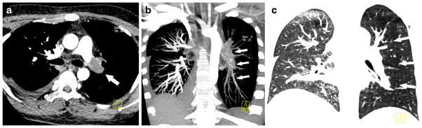
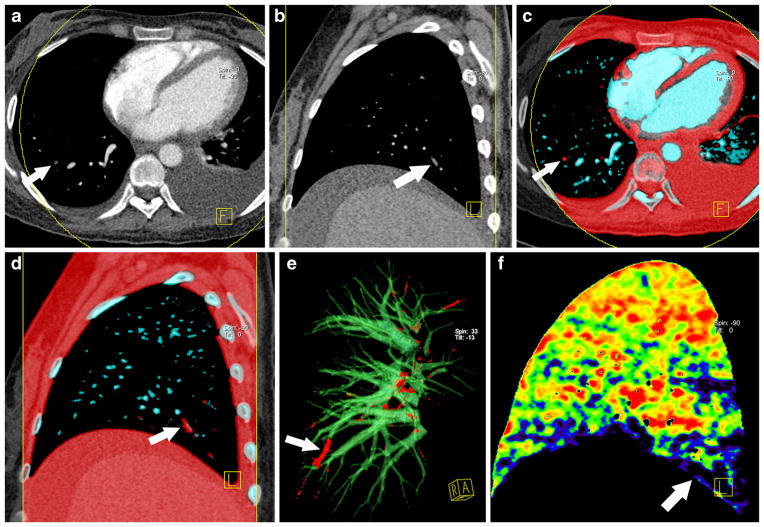
Pulmonary embolism is a life-threatening condition affecting people of all ages. Multidetector row CT pulmonary angiography has improved the imaging of pulmonary embolism in both adults and children and is now regarded as the routine modality for detection of pulmonary embolism. Advanced CT pulmonary angiography techniques developed in recent years, such as dual-energy CT, have been applied as a one-stop modality for pulmonary embolism diagnosis in children, as they can simultaneously provide anatomical and functional information. We discuss CT pulmonary angiography techniques, common and uncommon findings of pulmonary embolism in both conventional and dual-energy CT pulmonary angiography, and radiation dose considerations.


Conflict of interest statement

Conflicts of interest None

Figures

Fig. 1
Fig. 1
Complete occlusion of the left pulmonary artery in a 13-year-old boy with focal segmental glomerulosclerosis. a Contrast-enhanced axial CT image shows low-attenuation complete filling defect in left pulmonary artery and dilation of the artery (arrow). b Coronal maximum intensity projection image shows filling defect in left pulmonary arteries (arrows). c Coronal image with lung window shows the left lung oligemia caused by the large central pulmonary embolus (arrows)
Fig. 2
Fig. 2
Partial filling defects of multiple pulmonary arteries in a 15-year-old girl with systemic lupus erythematosus. ab Axial and coronal contrast-enhanced CT images demonstrate central filling defect in right pulmonary artery branch and eccentric partial filling defect in left pulmonary artery branch (arrows)
Fig. 3
Fig. 3
Dual-energy CT with vascular rendering in the detection of pulmonary embolus in a 14-year-old boy with podocytopathy. a–b Conventional CT images and (c–f) parametricized CT images from the same scan. Right lower pulmonary arteries are coded red (arrows in c–e) and the other pulmonary arteries are coded blue, which correspond to arrows in a–b in conventional CT pulmonary angiography. A perfusion defect is demonstrated with vascular rendering (arrow in f) in corresponding lung segment
Fig. 4
Fig. 4
Hampton’s hump in a 17-year-old girl with focal segmental glomerulosclerosis. a Axial contrast-enhanced CT image illustrates a pulmonary embolus (arrow) in the right pulmonary artery trunk and bilateral pleural effusion. b–c One month later, follow-up dual-energy CT images show a subpleural triangular lung infarct (Hampton’s hump; white arrows)
Fig. 5
Fig. 5
Common artifacts mimicking perfusion defects in dual-energy CT lung perfusion imaging in a 10-year-old boy with nephrotic syndrome. a–b Beam hardening artifacts and respiratory artifacts in dual-energy CT lung perfusion images. Radial beam hardening artifacts (thin white arrows) appear around vessels (thick white arrow) filled with contrast agent (a). Respiratory artifacts could be recognized in sagittal image as strip-shaped (white arrows) rather than wedge-shaped (yellow arrow in b)
Fig. 6
Fig. 6
Maximal diameter measurement of the right and left ventricles in axial contrast-enhanced CT image in a 15-year-old girl with podocytopathy. Measurements should be performed at the levels of right and left atrioventricular valve. Note the filling defects in the left inferior pulmonary arteries (arrow). RA right atrium, LA left atrium, RV right ventricle, LV left ventricle

References

    1. Tapson VF. Acute pulmonary embolism. N Engl J Med. 2008;358:1037–1052. - PubMed
    1. Ritchie G, McGurk S, McCreath C, et al. Prospective evaluation of unsuspected pulmonary embolism on contrast enhanced multidetector CT (MDCT) scanning. Thorax. 2007;62:536–540. - PMC - PubMed
    1. Victoria T, Mong A, Altes T, et al. Evaluation of pulmonary embolism in a pediatric population with high clinical suspicion. Pediatr Radiol. 2009;39:35–41. - PubMed
    1. Kritsaneepaiboon S, Lee EY, Zurakowski D, et al. MDCT pulmonary angiography evaluation of pulmonary embolism in children. AJR Am J Roentgenol. 2009;192:1246–1252. - PubMed
    1. Zhang LJ, Zhang Z, Li SJ, et al. Pulmonary embolism and renal vein thrombosis in patients with nephrotic syndrome: prospective evaluation of prevalence and risk factors with CT. Radiology. 2014;273:897–906. - PubMed

Publication types

LinkOut - more resources