Notch receptor expression in human brain arteriovenous malformations
- PMID: 25846406
- PMCID: PMC4549049
- DOI: 10.1111/jcmm.12580
Notch receptor expression in human brain arteriovenous malformations
Abstract
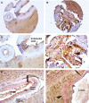
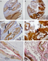
The roles of the Notch pathway proteins in normal adult vascular physiology and the pathogenesis of brain arteriovenous malformations are not well-understood. Notch 1 and 4 have been detected in human and mutant mice vascular malformations respectively. Although mutations in the human Notch 3 gene caused a genetic form of vascular stroke and dementia, its role in arteriovenous malformations development has been unknown. In this study, we performed immunohistochemistry screening on tissue microarrays containing eight surgically resected human brain arteriovenous malformations and 10 control surgical epilepsy samples. The tissue microarrays were evaluated for Notch 1-4 expression. We have found that compared to normal brain vascular tissue Notch-3 was dramatically increased in brain arteriovenous malformations. Similarly, Notch 4 labelling was also increased in vascular malformations and was confirmed by western blot analysis. Notch 2 was not detectable in any of the human vessels analysed. Using both immunohistochemistry on microarrays and western blot analysis, we have found that Notch-1 expression was detectable in control vessels, and discovered a significant decrease of Notch 1 expression in vascular malformations. We have demonstrated that Notch 3 and 4, and not Notch 1, were highly increased in human arteriovenous malformations. Our findings suggested that Notch 4, and more importantly, Notch 3, may play a role in the development and pathobiology of human arteriovenous malformations.
Keywords: BAVMs; cell signalling; endothelial cells; vascular malformations.
© 2015 The Authors. Journal of Cellular and Molecular Medicine published by John Wiley & Sons Ltd and Foundation for Cellular and Molecular Medicine.
Figures


References
-
- Al-Shahi R, Stapf C. The prognosis and treatment of arteriovenous malformations of the brain. Pract Neurol. 2005;5:194–205.
-
- Friedlander RM. Clinical practice. Arteriovenous malformations of the brain. N Engl J Med. 2007;356:2704–12. - PubMed
-
- Wang HU, Chen ZF, Anderson DJ. Molecular distinction and angiogenic interaction between embryonic arteries and veins revealed by ephrin-B2 and its receptor Eph-B4. Cell. 1998;93:741–53. - PubMed
-
- Bray SJ. Notch signalling: a simple pathway becomes complex. Nat Rev Mol Cell Bio. 2006;7:678–89. - PubMed
Publication types
MeSH terms
Substances
LinkOut - more resources
Full Text Sources
Other Literature Sources
Miscellaneous

