Activation of platelet protease-activated receptor-1 induces epithelial-mesenchymal transition and chemotaxis of colon cancer cell line SW620
- PMID: 25846512
- PMCID: PMC4431448
- DOI: 10.3892/or.2015.3897
Activation of platelet protease-activated receptor-1 induces epithelial-mesenchymal transition and chemotaxis of colon cancer cell line SW620
Erratum in
-
[Corrigendum] Activation of platelet protease-activated receptor-1 induces epithelial-mesenchymal transition and chemotaxis of colon cancer cell line SW620.Oncol Rep. 2016 Feb;35(2):1222. doi: 10.3892/or.2015.4438. Epub 2015 Nov 20. Oncol Rep. 2016. PMID: 26718652 Free PMC article.
Abstract
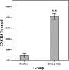
The aim of the present study was to examine the role of protease-activated receptor-1 (PAR1)-stimulated platelet activation in the epithelial-mesenchymal transition (EMT) and migration of colon cancer cells, and to identify the underlying mechanisms. TFLLR-NH2, a PAR1 agonist, was used to activate platelets and the platelet supernatants were used to treat the SW620 colon cancer cell line. Expression of E-cadherin and vimentin on SW620 cells was detected by immunofluorescence and western blotting, and the level of the transforming growth factor β1 (TGF-β1) was measured using ELISA following the activation of platelets by TFLLR-NH2. miR-200b expression was detected using quantitative PCR in SW620 cells. In order to investigate the chemotactic ability of the SW620 cells, the expression of CXC chemokine receptor type 4 (CXCR4) was measured by flow cytometry. Transwell migration assays were performed following exposure of the cells to the supernatant of PAR1-activated platelets. SW620 cells cultured in the supernatant of TFLLR-NH2-activated platelets upregulated E-cadherin expression and downregulated the vimentin expression. In the in vitro platelet culture system, a TFLLR-NH2 dose-dependent increase of secreted TGF-β1 was detected in the supernatant. The activation of PAR1 on the platelets led to the inhibition of miR-200b expression in the SW620 cells that were cultured in platelet-conditioned media. The number of SW620 cells that penetrated through the Transwell membrane increased with the dose of TFLLR-NH2 used to treat the platelets. The percentage of CXCR4-positive SW620 cells was significantly higher when they were exposed to the supernatant of platelets cultured for 24 h with PAR1 agonist than when cultured in non-conditioned media (40.89 ± 6.74 vs. 3.47 ± 1.40%, P < 0.01). Platelet activation with a PAR1 agonist triggered TGF-β secretion, which induced EMT of SW620 human colon cancer cells via the downregulation of miR-200b expression, and activated platelets had a chemotactic effect on colon cancer cells mediated by the upregulation of CXCR4 on the cell surface.
Figures


Similar articles
-
Prospero homeobox 1 promotes epithelial-mesenchymal transition in colon cancer cells by inhibiting E-cadherin via miR-9.Clin Cancer Res. 2012 Dec 1;18(23):6416-25. doi: 10.1158/1078-0432.CCR-12-0832. Epub 2012 Oct 8. Clin Cancer Res. 2012. PMID: 23045246
-
Resveratrol suppresses epithelial-to-mesenchymal transition in colorectal cancer through TGF-β1/Smads signaling pathway mediated Snail/E-cadherin expression.BMC Cancer. 2015 Mar 5;15:97. doi: 10.1186/s12885-015-1119-y. BMC Cancer. 2015. PMID: 25884904 Free PMC article.
-
Stereospecific effects of ginsenoside 20-Rg3 inhibits TGF-β1-induced epithelial-mesenchymal transition and suppresses lung cancer migration, invasion and anoikis resistance.Toxicology. 2014 Aug 1;322:23-33. doi: 10.1016/j.tox.2014.04.002. Epub 2014 May 2. Toxicology. 2014. PMID: 24793912
-
Danshensu Attenuated Epithelial-Mesenchymal Transformation and Chemoresistance of Colon Cancer Cells Induced by Platelets.Front Biosci (Landmark Ed). 2022 May 17;27(5):160. doi: 10.31083/j.fbl2705160. Front Biosci (Landmark Ed). 2022. PMID: 35638427
-
Novel insights into the regulation of cyclooxygenase-2 expression by platelet-cancer cell cross-talk.Biochem Soc Trans. 2015 Aug;43(4):707-14. doi: 10.1042/BST20140322. Epub 2015 Aug 3. Biochem Soc Trans. 2015. PMID: 26551717 Free PMC article. Review.
Cited by
-
Platelets involved tumor cell EMT during circulation: communications and interventions.Cell Commun Signal. 2022 Jun 3;20(1):82. doi: 10.1186/s12964-022-00887-3. Cell Commun Signal. 2022. PMID: 35659308 Free PMC article. Review.
-
Signaling Crosstalk of TGF-β/ALK5 and PAR2/PAR1: A Complex Regulatory Network Controlling Fibrosis and Cancer.Int J Mol Sci. 2018 May 24;19(6):1568. doi: 10.3390/ijms19061568. Int J Mol Sci. 2018. PMID: 29795022 Free PMC article. Review.
-
Platelet secretion in inflammatory and infectious diseases.Platelets. 2017 Mar;28(2):155-164. doi: 10.1080/09537104.2016.1240766. Epub 2016 Nov 16. Platelets. 2017. PMID: 27848259 Free PMC article. Review.
-
Aspirin and cancer: biological mechanisms and clinical outcomes.Open Biol. 2022 Sep;12(9):220124. doi: 10.1098/rsob.220124. Epub 2022 Sep 14. Open Biol. 2022. PMID: 36099932 Free PMC article. Review.
-
Protease-activated receptor-1 (PAR-1): a promising molecular target for cancer.Oncotarget. 2017 Sep 18;8(63):107334-107345. doi: 10.18632/oncotarget.21015. eCollection 2017 Dec 5. Oncotarget. 2017. PMID: 29291033 Free PMC article. Review.
References
-
- Thiery JP. Epithelial-mesenchymal transitions in cancer onset and progression. Bull Acad Natl Med. 2009;193:1969–1979. In French. - PubMed
Publication types
MeSH terms
Substances
LinkOut - more resources
Full Text Sources
Other Literature Sources

