

Age-related sensitivity to endotoxin-induced liver inflammation: Implication of inflammasome/IL-1β for steatohepatitis
- PMID: 25847140
- PMCID: PMC4531067
- DOI: 10.1111/acel.12305
Age-related sensitivity to endotoxin-induced liver inflammation: Implication of inflammasome/IL-1β for steatohepatitis
Abstract
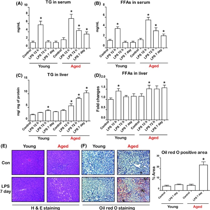
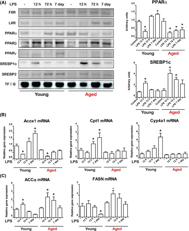
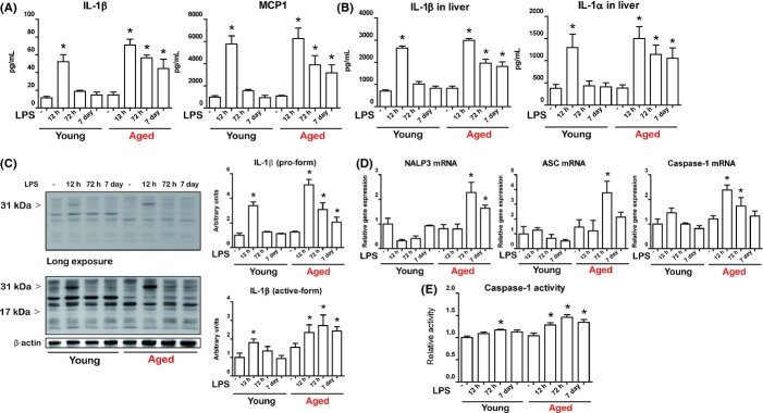
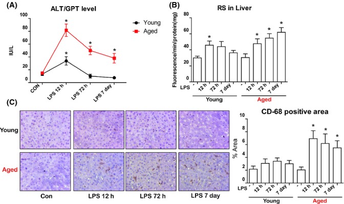
Aging is associated with increased vulnerability to inflammatory challenge. However, the effects of altered inflammatory response on the metabolic status of tissues or organs are not well documented. In this study, we present evidence demonstrating that lipopolysaccharide (LPS)-induced upregulation of the inflammasome/IL-1β pathway is accompanied with an increased inflammatory response and abnormal lipid accumulation in livers of aged rats. To monitor the effects of aging on LPS-induced inflammation, we administered LPS (2 mg kg(-1) ) to young (6-month old) and aged (24-month old) rats and found abnormal lipid metabolism in only aged rats with increased lipid accumulation in the liver. This lipid accumulation in the liver was due to the dysregulation of PPARα and SREBP1c. We also observed severe liver inflammation in aged rats as indicated by increased ALT levels in serum and increased Kupffer cells in the liver. Importantly, among many inflammation-associated factors, the aged rat liver showed chronically increased IL-1β production. Increased levels of IL-1β were caused by the upregulation of caspase-1 activity and inflammasome activation. In vitro studies with HepG2 cells demonstrated that treatment with IL-1β significantly induced lipid accumulation in hepatocytes through the regulation of PPARα and SREBP1c. In summary, we demonstrated that LPS-induced liver inflammation and lipid accumulation were associated with a chronically overactive inflammasome/IL-1β pathway in aged rat livers. Based on the present findings, we propose a mechanism of aging-associated progression of steatohepatitis induced by endotoxin, delineating a pathogenic role of the inflammasome/IL-1β pathway involved in lipid accumulation in the liver.
Keywords: IL-1β; LPS; aging; inflammasome; inflammation; lipid accumulation.
© 2015 The Authors. Aging Cell published by the Anatomical Society and John Wiley & Sons Ltd.
Figures


Similar articles
-
Hepatic interleukin-6 production is maintained during endotoxin tolerance and facilitates lipid accumulation.Immunobiology. 2017 Jun;222(6):786-796. doi: 10.1016/j.imbio.2017.01.003. Epub 2017 Jan 20. Immunobiology. 2017. PMID: 28132721
-
Peroxisome proliferator-activated receptor-delta agonist ameliorated inflammasome activation in nonalcoholic fatty liver disease.World J Gastroenterol. 2015 Dec 7;21(45):12787-99. doi: 10.3748/wjg.v21.i45.12787. World J Gastroenterol. 2015. PMID: 26668503 Free PMC article.
-
Mechanisms that lead to the regulation of NLRP3 inflammasome expression and activation in human dental pulp fibroblasts.Mol Immunol. 2015 Aug;66(2):253-62. doi: 10.1016/j.molimm.2015.03.009. Epub 2015 Apr 2. Mol Immunol. 2015. PMID: 25863775
-
Inflammasome activation in the liver: Focus on alcoholic and non-alcoholic steatohepatitis.Clin Res Hepatol Gastroenterol. 2015 Sep;39 Suppl 1:S18-23. doi: 10.1016/j.clinre.2015.06.012. Epub 2015 Jul 26. Clin Res Hepatol Gastroenterol. 2015. PMID: 26216030 Review.
-
IL-18 and IL-1β Gene Polymorphisms: The Plausible Risk Factors for Chronic Hepatitis B.Viral Immunol. 2019 Jun;32(5):208-213. doi: 10.1089/vim.2018.0155. Epub 2019 May 14. Viral Immunol. 2019. PMID: 31084469 Review.
Cited by
-
FoxO6-mediated IL-1β induces hepatic insulin resistance and age-related inflammation via the TF/PAR2 pathway in aging and diabetic mice.Redox Biol. 2019 Jun;24:101184. doi: 10.1016/j.redox.2019.101184. Epub 2019 Apr 3. Redox Biol. 2019. PMID: 30974318 Free PMC article.
-
Effect of Oleoylethanolamide-Based Dietary Supplement on Systemic Inflammation in the Development of Alimentary-Induced Obesity in Mice.Nutrients. 2023 Oct 12;15(20):4345. doi: 10.3390/nu15204345. Nutrients. 2023. PMID: 37892420 Free PMC article.
-
Medicinal Mushroom Extracts from Hericium coralloides and Trametes versicolor Exert Differential Immunomodulatory Effects on Immune Cells from Older Adults In Vitro.Nutrients. 2023 May 8;15(9):2227. doi: 10.3390/nu15092227. Nutrients. 2023. PMID: 37432355 Free PMC article.
-
Modelling physical resilience in ageing mice.Mech Ageing Dev. 2019 Jan;177:91-102. doi: 10.1016/j.mad.2018.10.001. Epub 2018 Oct 2. Mech Ageing Dev. 2019. PMID: 30290161 Free PMC article. Review.
-
Organ-differential Roles of Akt/FoxOs Axis as a Key Metabolic Modulator during Aging.Aging Dis. 2021 Oct 1;12(7):1713-1728. doi: 10.14336/AD.2021.0225. eCollection 2021 Oct. Aging Dis. 2021. PMID: 34631216 Free PMC article. Review.
References
-
- Affò S, Morales-Ibanez O, Rodrigo-Torres D, Altamirano J, Blaya D, Dapito DH, Millán C, Coll M, Caviglia JM, Arroyo V, Caballería J, Schwabe RF, Ginès P, Bataller R, Sancho-Bru P. CCL20 mediates lipopolysaccharide induced liver injury and is a potential driver of inflammation and fibrosis in alcoholic hepatitis. Gut. 2014;63:1782–1792. - PMC - PubMed
-
- Carré JE, Singer M. Cellular energetic metabolism in sepsis: the need for a systems approach. Biochim. Biophys. Acta. 2008;1777:763–771. - PubMed
-
- Chen X, Zhang C, Zhao M, Shi CE, Zhu RM, Wang H, Zhao H, Wei W, Li JB, Xu DX. Melatonin alleviates lipopolysaccharide-induced hepatic SREBP-1c activation and lipid accumulation in mice. J. Pineal Res. 2011;51:416–425. - PubMed
-
- Desvergne B, Michalik L, Wahli W. Transcriptional regulation of metabolism. Physiol. Rev. 2006;86:465–514. - PubMed
-
- Dorshkind K, Montecino-Rodriguez E, Signer RA. The ageing immune system: is it ever too old to become young again? Nat. Rev. Immunol. 2009;9:57–62. - PubMed
Publication types
MeSH terms
Substances
LinkOut - more resources
Full Text Sources
Other Literature Sources
Medical
