. 2015 Jun 5;290(23):14407-17.
doi: 10.1074/jbc.M115.653600. Epub 2015 Apr 6.

Mycobacterium tuberculosis TlyA Protein Negatively Regulates T Helper (Th) 1 and Th17 Differentiation and Promotes Tuberculosis Pathogenesis

Affiliations

Mycobacterium tuberculosis TlyA Protein Negatively Regulates T Helper (Th) 1 and Th17 Differentiation and Promotes Tuberculosis Pathogenesis

Md Aejazur Rahman et al. J Biol Chem. .

Abstract

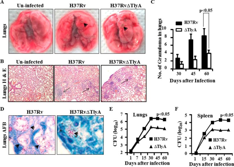
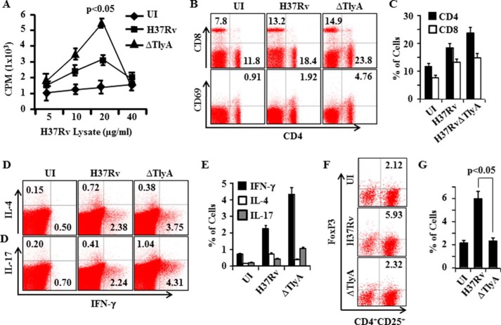
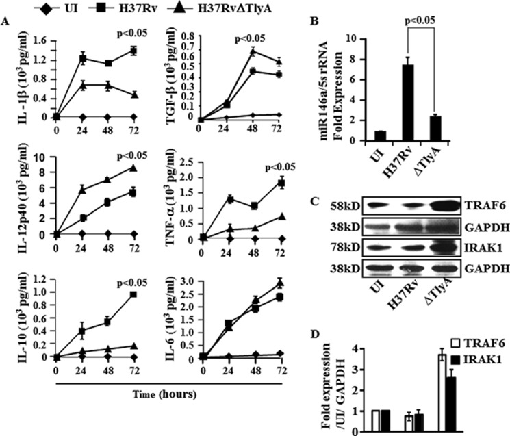
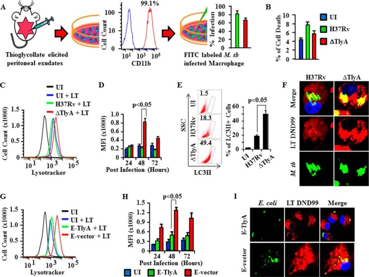
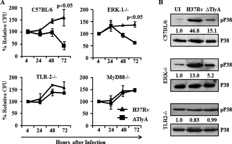
Mycobacterium tuberculosis, the causative agent of tuberculosis, is an ancient pathogen and a major cause of death worldwide. Although various virulence factors of M. tuberculosis have been identified, its pathogenesis remains incompletely understood. TlyA is a virulence factor in several bacterial infections and is evolutionarily conserved in many Gram-positive bacteria, but its function in M. tuberculosis pathogenesis has not been elucidated. Here, we report that TlyA significantly contributes to the pathogenesis of M. tuberculosis. We show that a TlyA mutant M. tuberculosis strain induces increased IL-12 and reduced IL-1β and IL-10 cytokine responses, which sharply contrasts with the immune responses induced by wild type M. tuberculosis. Furthermore, compared with wild type M. tuberculosis, TlyA-deficient M. tuberculosis bacteria are more susceptible to autophagy in macrophages. Consequently, animals infected with the TlyA mutant M. tuberculosis organisms exhibited increased host-protective immune responses, reduced bacillary load, and increased survival compared with animals infected with wild type M. tuberculosis. Thus, M. tuberculosis employs TlyA as a host evasion factor, thereby contributing to its virulence.

Keywords: Mycobacterium tuberculosis; T helper cells; cytokine; vaccine; virulence factor.


Figures

FIGURE 1.
FIGURE 1.
General analysis of lungs and spleen of H37Rv- and H37RvΔTlyA-challenged BALB/c mice. BALB/c mice were challenged with H37Rv or H37RvΔTlyA by the aerosol route, and lungs and spleen of the infected mice were harvested at different time points. A, gross pictures of lungs of uninfected, H37Rv-infected, and H37RvΔTlyA-infected mice. B, histology of the lung tissue sections at 45 days post-infection stained with hemotoxylin & eosin. C, numbers of granulomas in lungs at different time points after infection. D, acid fast staining of bacilli. E, colony-forming units from the lung homogenates of H37Rv- and H37RvΔTlyA-infected mice at different time points. F, colony-forming units from splenocytes of mice that were infected with H37Rv or the H37RvΔTlyA mutant. The results shown are representative of three independent experiments with five mice per group per time point.
FIGURE 2.
FIGURE 2.
T cell proliferation and FACS analyses of T cell subsets. A, T cell proliferation from spleen of H37Rv- and H37RvΔTlyA-infected mice. B and C, FACS analysis shows the percentage of CD4+T cells, CD8+T cells, and activation marker CD69- and CD25-positive cells in M. tuberculosis-infected mice. D and E, T cells secreting IFN-γ, IL-4, or IL-17 among splenocytes of M. tuberculosis-infected mice. F and G, percentage of Treg cells (FoxP3+ CD4+CD25+cells) among splenocytes of M. tuberculosis-infected mice. The results shown are representative of three independent experiments with three mice per group per time point. UI, uninfected.
FIGURE 3.
FIGURE 3.
Cytokine profiling of H37RvΔTlyA-infected dendritic cells. A, DCs of mice were infected with H37Rv or H37RvΔTlyA, and cytokines from culture supernatant were assayed at different time points and compared with uninfected (UI) DCs. Luminex assay showing the cytokine concentration of IL-1β, IL-10, IL-12p40, IL-6, TGF-β, and TNF-α in the culture supernatants of H37Rv-infected (■) or H37RvΔTlyA-infected (▴) DCs compared with uninfected DCs (♦). The results shown are representative of at least three independent experiments with three replicates. B, miR146a expression in H37Rv- and H37RvΔTlyA-infected DCs after 48 h of infection. The results shown are representative of at least three independent experiments with three replicates. C and D, total cell lysates from uninfected or M. tuberculosis (H37Rv or H37RvΔTlyA)-infected macrophages were electrophoresed on 12% SDS-polyacrylamide gels and transferred onto nitrocellulose membranes. These membranes were probed with anti-TRAF-6 antibodies, then stripped and reprobed with anti-GAPDH as an equal loading control. Immunoblots are representative of three independent experiments.
FIGURE 4.
FIGURE 4.
TlyA induces autophagy in M. tuberculosis (M. tb.)-infected macrophages. A, schematic model to show the purity of macrophages obtained from thioglycollate-elicited peritoneal exudates and the percentage of infected macrophages using FITC-labeled H37Rv and H37RvΔTlyA, as revealed by flow cytometry. B, cell death after 72 h was assessed by propidium iodide staining followed by flow cytometry. C, autophagy was assessed by measurement of LysoTracker Red DND-99 using flow cytometry. FITC-labeled H37Rv- and H37RvΔTlyA-infected macrophages were stained with DND-99 and analyzed on channel APC-A. Representative histograms are displayed. Data overlays were performed by FlowJo. The y axis shows cell counts, and the x axis represents fluorescence intensity in log scale and shifting of DND-99. D, mean fluorescence intensity (MFI) value of autophagy. E, flow cytometry data to show the percentage of LC3II expression. F, confocal microscopy of FITC-labeled H37Rv- and H37RvΔTlyA-infected macrophages. Infected macrophages were stained with DND-99 and viewed under a ×100 optical zoom. G, autophagy was assessed by measurement of LysoTracker Red DND-99 using flow cytometry. Macrophages infected with FITC-labeled TlyA-expressing E. coli (E-TlyA) or mock vector transformed E. coli (E-vector) were stained with DND-99 and analyzed on channel APC-A. Representative histograms are displayed. Data overlays were performed by FlowJo. The y axis shows cell counts, and the x axis represents fluorescence intensity in log scale and shifting of DND-99. H, mean fluorescence intensity value of autophagy. I, confocal microscopy of FITC-labeled E-TlyA- and E-vector-infected macrophages. Infected macrophages were stained with LysoTracker-DND-99 and viewed under a ×100 optical zoom. The results shown are representative of three independent experiments. UI, uninfected.
FIGURE 5.
FIGURE 5.
TlyA activates p38-MAPK in the TLR2-MyD88 signaling pathway. A, macrophages from wild type C57BL/6 and ERK−/−, TLR2−/−, and MyD88−/− mice were infected with H37Rv or H37RvΔTlyA to determine the relative colony-forming units at different time points. The relative colony-forming units of H37RvΔTlyA was decreased in macrophages isolated from wild type and ERK−/− mice at 72 h post-infection, compared with H37Rv. Moreover, colony-forming units of H37Rv and H37RvΔTlyA were similar to wild type in macrophages isolated from TLR2−/− and MyD88−/− mice. B, Western blot analysis for phospho-p38 in macrophages isolated from wild type C57BL/6 and ERK−/− and TLR2−/− mice infected with H37Rv or H37RvΔTlyA at 24 h post-infection. H37Rv-infected macrophages from wild type and ERK−/− mice showed activation of p38-MAPK compared with uninfected (UI) and H37RvΔTlyA-infected macrophages. In contrast, p38-MAPK was equally activated in TLR2−/− and wild type mice for uninfected and H37Rv-or H37RvΔTlyA-infected macrophages. The results shown are representative of three independent experiments with three replicates.

Similar articles

Cited by

References

    1. Dye C. (2006) Global epidemiology of tuberculosis. Lancet 367, 938–940 - PubMed
    1. Ehrt S., Schnappinger D. (2009) Mycobacterial survival strategies in the phagosome: defence against host stresses. Cell. Microbiol. 11, 1170–1178 - PMC - PubMed
    1. Smith I. (2003) Mycobacterium tuberculosis pathogenesis and molecular determinants of virulence. Clin. Microbiol. Rev. 16, 463–496 - PMC - PubMed
    1. Glickman M. S., Jacobs W. R., Jr. (2001) Microbial pathogenesis of Mycobacterium tuberculosis: dawn of a discipline. Cell 104, 477–485 - PubMed
    1. Ahmad S. (2011) Pathogenesis, immunology, and diagnosis of latent Mycobacterium tuberculosis infection. Clin. Dev. Immunol. 2011, 814943. - PMC - PubMed

MeSH terms