. 2015 May 22;290(21):13479-89.
doi: 10.1074/jbc.M114.636050. Epub 2015 Apr 6.

PRMT1 Is a Novel Regulator of Epithelial-Mesenchymal-Transition in Non-small Cell Lung Cancer

Affiliations

PRMT1 Is a Novel Regulator of Epithelial-Mesenchymal-Transition in Non-small Cell Lung Cancer

Sreedevi Avasarala et al. J Biol Chem. .

Abstract

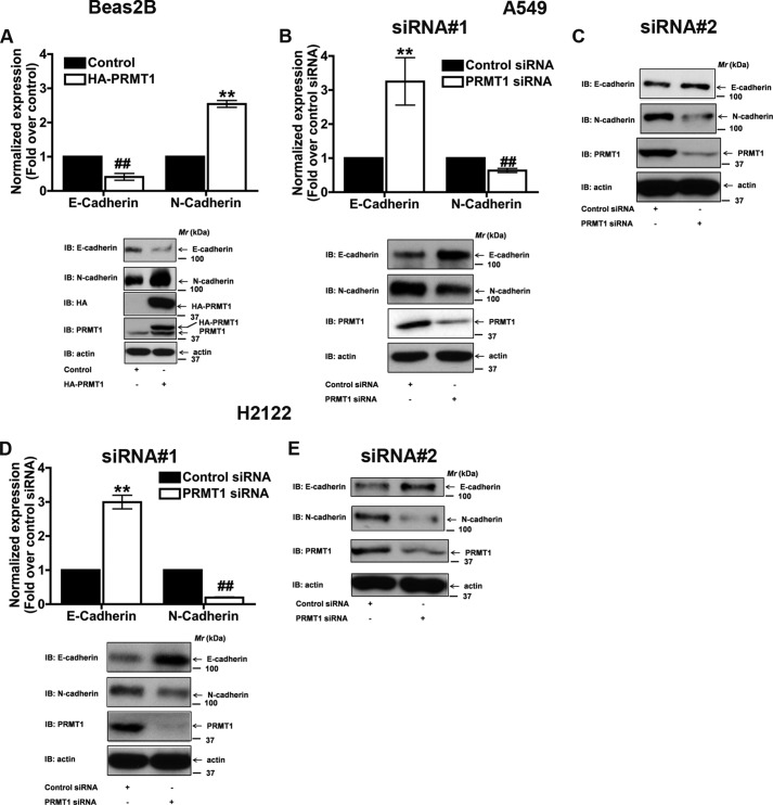
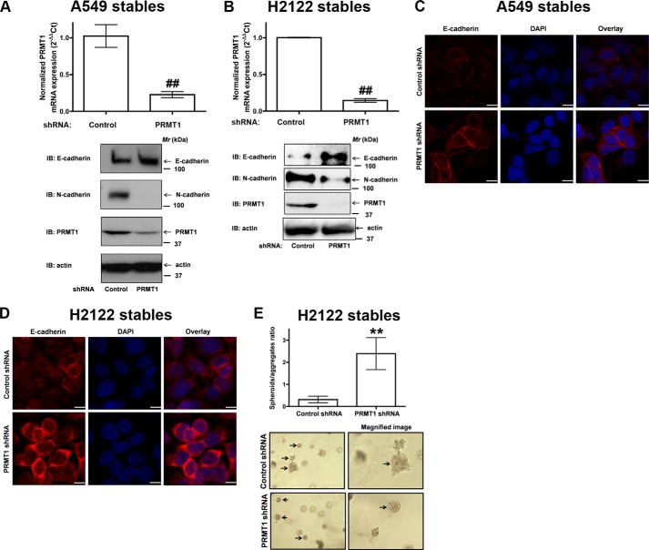
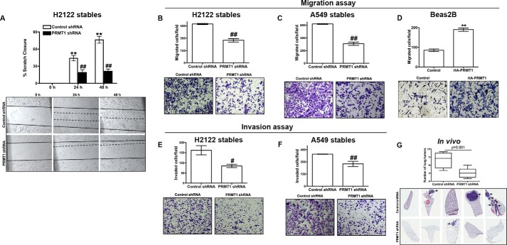
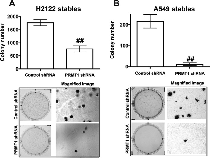
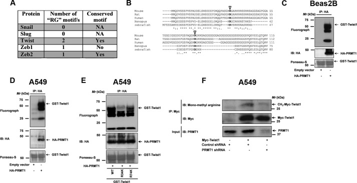
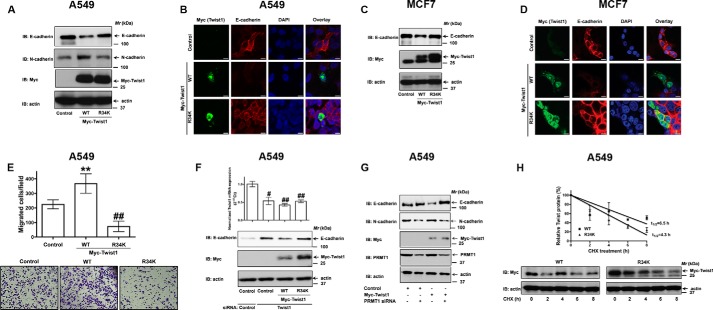
Protein arginine methyl transferase 1 (PRMT1) was shown to be up-regulated in cancers and important for cancer cell proliferation. However, the role of PRMT1 in lung cancer progression and metastasis remains incompletely understood. In the present study, we show that PRMT1 is an important regulator of epithelial-mesenchymal transition (EMT), cancer cell migration, and invasion, which are essential processes during cancer progression, and metastasis. Additionally, we have identified Twist1, a basic helix-loop-helix transcription factor and a well-known E-cadherin repressor, as a novel PRMT1 substrate. Taken together, we show that PRMT1 is a novel regulator of EMT and arginine 34 (Arg-34) methylation of Twist1 as a unique "methyl arginine mark" for active E-cadherin repression. Therefore, targeting PRMT1-mediated Twist1 methylation might represent a novel strategy for developing new anti-invasive/anti-metastatic drugs. Moreover, methylated Twist1 (Arg-34), as such, could also emerge as a potential important biomarker for lung cancer.

Keywords: N-cadherin; Prmt1; Twist1; cadherin-1 (CDH1) (epithelial cadherin) (E-cadherin); epithelial-mesenchymal transition (EMT); metastasis; protein methylation.


Figures

FIGURE 1.
FIGURE 1.
PRMT1 is a novel regulator of EMT. A, overexpression of PRMT1 in a non-transformed lung epithelial cell line (Beas2B) induced EMT, as detected by a decrease in E-cadherin and an increase in N-cadherin. Treatment of A549 (B and C) or H2122 (D and E) cells with two different PRMT1 siRNA sequences (siRNA#1 (Invitrogen), and siRNA#2 (Santa Cruz Biotechnology)) reversed EMT, as defined by an increase in E-cadherin and a decrease in N-cadherin. E-cadherin and N-cadherin protein levels were quantified by densitometry, normalized to their corresponding actin controls, and are represented in the figures as fold changes over control. **, p < 0.01; or ##, p < 0.01; versus control.
FIGURE 2.
FIGURE 2.
PRMT1 knockdown induce epithelial phenotype in NSCLC cells. A549 (A) or H2122 (B) clones with stable expression of control shRNA or PRMT1 shRNA were generated as described under “Experimental Procedures.” Upper panel represents qPCR analysis for PRMT1 knockdown efficiency, whereas lower panels represent immunoblots with E-cadherin, N-cadherin, PRMT1, and actin antibodies. ##, p < 0.01; versus control shRNA. C and D, A549, and H2122 clones stably expressing either control shRNA or PRMT1 shRNA were fixed, permeabilized and immunostained with anti-E-cadherin antibodies, and the expression of E-cadherin was visualized by indirect immunofluorescence and confocal microscopy. Scale bar 10 μm. E, H2122 clones stably expressing either control shRNA or PRMT1 shRNA were embedded in Matrigel as single cells as described under “Experimental Procedures.” The upper panel represents the ratio of spheroids/aggregates, whereas representative low power and magnified images are presented in the lower panel. **, p < 0.01; versus control shRNA.
FIGURE 3.
FIGURE 3.
PRMT1 knockdown results in reduced cell migration and invasion. A, A 3-mm scrape wound was created in confluent cultures of H2122 clones with stable expression of either control shRNA or PRMT1 shRNA, and cell migration was recorded as described under “Experimental Procedures.” The upper panel represents the quantification of migration as described under “Experimental Procedures,” whereas representative images were presented in the lower panel. B and C, migration of H2122 (B) or A549 (C) clones with stable expression of either control shRNA or PRMT1 shRNA were assayed in trans-well inserts as described under “Experimental Procedures.” Top panel represents the number of cells migrated, whereas representative images are displayed in the lower panel. D, migration of Beas2B cells transiently transfected with empty vector or HA-tagged PRMT1 were assayed in trans-well inserts as described in the methods. Top panel represents the number of cells migrated, whereas representative images are displayed in the lower panel. **, p < 0.01; versus control. E and F, invasion of H2122 (E) or A549 (F) clones with stable expression of either control shRNA or PRMT1 shRNA were assayed in trans-well inserts coated with Matrigel as described under “Experimental Procedures.” Top panel represents the number of cells invaded, whereas representative images are displayed in the lower panel. ##, p < 0.01; #, p < 0.05; versus control shRNA. G, H2122 clones stably expressing either control shRNA or PRMT1 shRNA were injected into athymic nude mice via tail-veins (n = 5). After 8 weeks, mice were sacrificed, visible tumors were counted and are represented in the graph, whereas representative H&E-stained lung sections were displayed in the lower panel.
FIGURE 4.
FIGURE 4.
PRMT1 regulates anchorage-independent growth. Anchorage-independent growth of H2122 (A) or A549 (B) clones with stable expression of either control shRNA or PRMT1 shRNA were assayed using soft agar assays as described under “Experimental Procedures.” Upper panel represents the number of colonies formed, whereas representative low power, and magnified images are displayed in the lower panel. ##, p < 0.01; versus control shRNA.
FIGURE 5.
FIGURE 5.
Twist1 is a novel PRMT1 substrate. A, In silico analysis of primary amino acid sequences of known E-cadherin repressors for PRMT methylation motifs. B, multiple sequence alignments of mouse, rat, human, Xenopus, and Zebrafish Twist1 amino acid sequences. Potential PRMT methylation sites are highlighted with open arrowheads. PRMT-mediated Twist1 methylation was evaluated by using an in vitro methylation reaction that includes HA-tagged PRMT1 purified either from Beas2B (C) or A549 (D) cell lysates, along with GST-Twist1, purified from E. coli and [3H]SAMe as a methyl donor as described under “Experimental Procedures.” The methylation status of Twist1 was revealed by SDS-PAGE followed by fluorography. After fluorography, the blots were stained with ponseau S to determine equal loading of the substrate (GST-Twist1, lower panel). E, PRMT1 purified from A549 cells were employed in an in vitro methylation reaction along with GST-Twist1 (wild-type) or its mutants R34K, and R74K and [3H]SAMe as a methyl donor. The methylation status of Twist1 and its mutants were later revealed by SDS-PAGE followed by fluorography. After fluorography, the blots were stained with Ponseau S to determine equal loading of the substrate (GST-Twist1 or its mutants, lower panel). F, to determine if Twist1 was methylated in vivo, A549 cells were co-transfected with Myc-tagged Twist1 and either control shRNA or PRMT1 shRNA. The lysates were later employed in immunoprecipiations with anti-myc antibodies. The methylation status of Twist1 was later determined by probing the blots with anti-monomethyl arginine-specific antibodies.
FIGURE 6.
FIGURE 6.
PRMT-mediated Twist1 methylation is required for E-cadherin repression. A, A549 cells were transiently transfected with either Myc-tagged wild-type or R34K mutant of Twist1. The lysates were later probed for the expression of E-cadherin and N-cadherin via immunoblotting with specific antibodies. B, A549 cells transfected with empty vector, Myc-tagged wild-type or R34K mutant of Twist1 were fixed, permeabilized, and co-immunostained with anti-myc antibodies and anti-E-cadherin antibodies, and the expressions of E-cadherin and Twist1 (Myc) were visualized by indirect immunofluorescence and confocal microscopy. Scale bar 5 μm. C, MCF7 cells were transiently transfected with either Myc-tagged wild-type or R34K mutant of Twist1. The lysates were later probed for the expression of E-cadherin and N-cadherin via immunoblotting with specific antibodies. D, MCF7 cells transfected with empty vector, Myc-tagged wild-type or R34K mutant of Twist1 were fixed, permeabilized and co-immunostained with anti-myc antibodies and anti-E-cadherin antibodies, and the expressions of E-cadherin and Twist1 (Myc) were visualized by indirect immunofluorescence and confocal microscopy. Scale bar 5 μm. E, migration of A549 cells transfected with empty vector, Myc-tagged wild-type or R34K mutant of Twist1 were determined using trans-well assays as described under “Experimental Procedures.” Top panel represents the number of cells migrated, whereas representative images are displayed in the lower panel. ##, p < 0.01; **, p < 0.01; versus control. F, A549 cells were co-transfected with Twist1 siRNA and either wild-type or R34K mutant of Twist1. The lysates were later probed for the expression of E-cadherin, Myc-Twist1 with specific antibodies. G, A549 cells were co-transfected with Myc-tagged Twist1 and either control siRNA or PRMT1 siRNA. Total lysates were later probed for the expression of E-cadherin, N-cadherin, PRMT1, and Twist1 (Myc). H, A549 cells transiently transfected with either Myc-tagged Twist1 or R34K mutant were treated with cycloheximide (100 μg/ml) for indicated periods of time, followed by immunoblotting and densitometric scanning. Upper panel represents the normalized Twist1 (Myc)/actin levels, whereas representative images are displayed in the lower panel.

Similar articles

Cited by

References

    1. Yang Y., Bedford M. T. (2013) Protein arginine methyltransferases and cancer. Nature Reviews. Cancer 13, 37–50 - PubMed
    1. Cha B., Jho E. H. (2012) Protein arginine methyltransferases (PRMTs) as therapeutic targets. Exp. Opin. Therap. Targets 16, 651–664 - PubMed
    1. Paik W. K., Kim S. (1968) Protein methylase I. Purification and properties of the enzyme. J. Biol. Chem. 243, 2108–2114 - PubMed
    1. Lin W. J., Gary J. D., Yang M. C., Clarke S., Herschman H. R. (1996) The mammalian immediate-early TIS21 protein and the leukemia-associated BTG1 protein interact with a protein-arginine N-methyltransferase. J. Biol. Chem. 271, 15034–15044 - PubMed
    1. Boffa L. C., Karn J., Vidali G., Allfrey V. G. (1977) Distribution of NG, NG,-dimethylarginine in nuclear protein fractions. Biochem. Biophys. Res. Commun. 74, 969–976 - PubMed

Publication types

MeSH terms