

Ductal metaplasia in oesophageal submucosal glands is associated with inflammation and oesophageal adenocarcinoma
- PMID: 25847432
- PMCID: PMC4592376
- DOI: 10.1111/his.12707
Ductal metaplasia in oesophageal submucosal glands is associated with inflammation and oesophageal adenocarcinoma
Abstract
Aims: Recent studies have suggested that oesophageal submucosal gland (ESMG) ducts harbour progenitor cells that may contribute to oesophageal metaplasia. Our objective was to determine whether histological differences exist between the ESMGs of individuals with and without oesophageal adenocarcinoma (EAC).
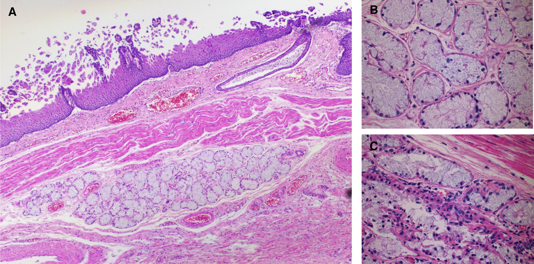
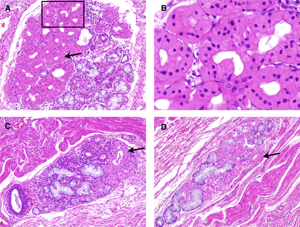
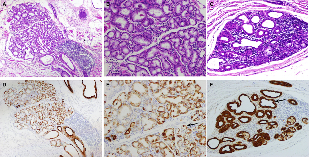
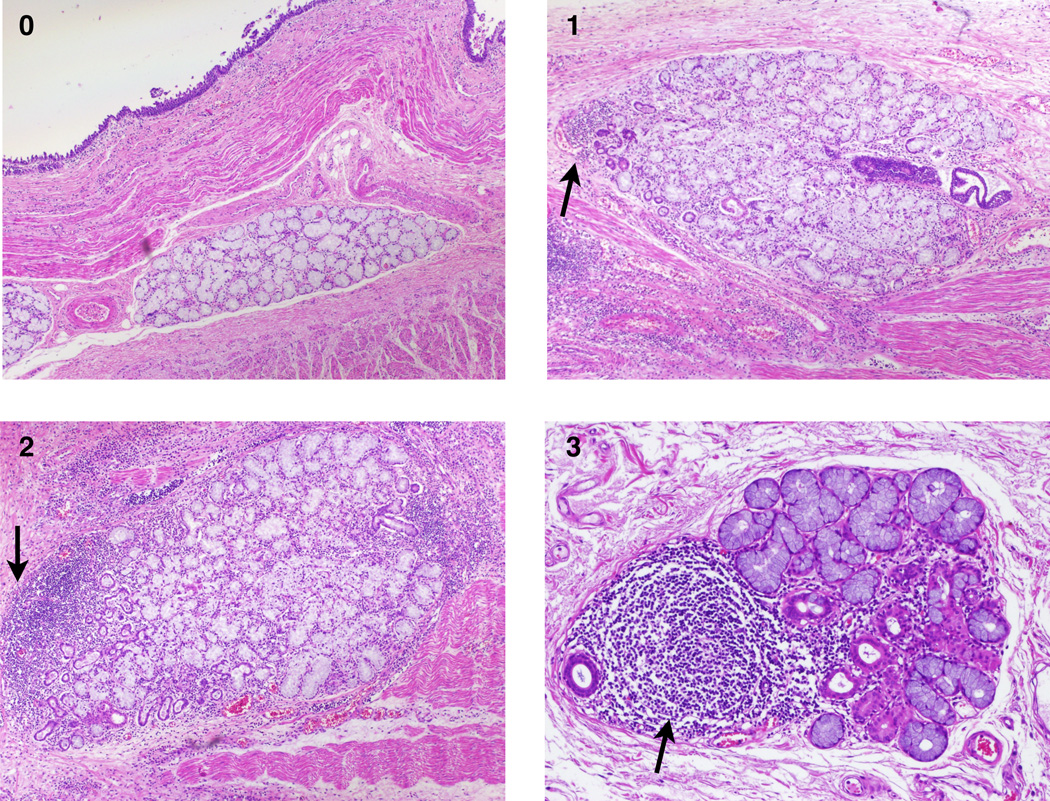
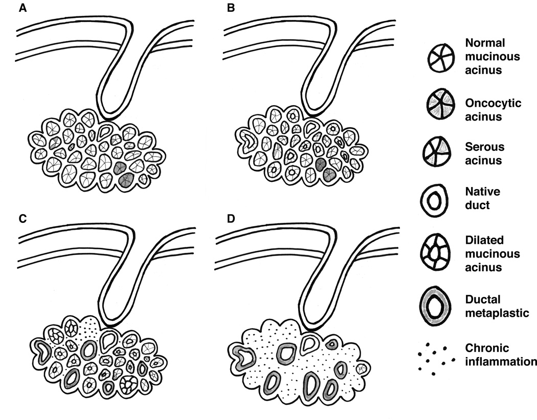
Methods and results: We performed histological assessment of 343 unique ESMGs from 30 control patients, 24 patients with treatment-naïve high-grade columnar dysplasia (HGD) or EAC, and 23 non-EAC oesophagectomy cases. A gastrointestinal pathologist assessed haematoxylin and eosin-stained ESMG images by using a scoring system that assigns individual ESMG acini to five histological types (mucous, serous, oncocytic, dilated, or ductal metaplastic). In our model, ductal metaplastic acini were more common in patients with HGD/EAC (12.7%) than in controls (3.5%) (P = 0.006). We also identified greater proportions of acini with dilation (21.9%, P < 0.001) and, to a lesser extent, ductal metaplasia (4.3%, P = 0.001) in non-EAC oesophagectomy cases than in controls. Ductal metaplasia tended to occur in areas of mucosal ulceration or tumour.
Conclusions: We found a clear association between ductal metaplastic ESMG acini and HGD/EAC. Non-EAC cases had dilated acini and some ductal dilation. Because ESMGs and ducts harbour putative progenitor cells, these associations could have significance for understanding the pathogenesis of EAC.
Keywords: Barrett's oesophagus; ductal metaplasia; oesophageal neoplasm; oesophageal submucosal gland; oesophagus.
© 2015 John Wiley & Sons Ltd.
Conflict of interest statement
Conflicts of Interest Statement: None of the authors have conflicts of interest to report.
Figures
References
-
- Pohl H, Welch HG. The role of overdiagnosis and reclassification in the marked increase of esophageal adenocarcinoma incidence. J Natl Cancer Inst. 2005;97:142–146. - PubMed
-
- Howlader N, Noone AM, Krapcho M, et al. Seer cancer statistics review. Bethesda, MD: National Cancer Institute; 2011. based upon November 2010 SEER data submission.
-
- Spechler SJ, Sharma P, Souza RF, Inadomi JM, Shaheen NJ. American gastroenterological association medical position statement on the management of barrett's esophagus. Gastroenterology. 2011;140:1084–1091. - PubMed
-
- Dulai GS, Guha S, Kahn KL, Gornbein J, Weinstein WM. Preoperative prevalence of barrett's esophagus in esophageal adenocarcinoma: A systematic review. Gastroenterology. 2002;122:26–33. - PubMed
-
- Hvid-Jensen F, Pedersen L, Drewes AM, Sorensen HT, Funch-Jensen P. Incidence of adenocarcinoma among patients with barrett's esophagus. N Engl J Med. 2011;365:1375–1383. - PubMed
Publication types
MeSH terms
Grants and funding
LinkOut - more resources
Full Text Sources
Other Literature Sources
Medical
