

Combination of 12-O-tetradecanoylphorbol-13-acetate with diethyldithiocarbamate markedly inhibits pancreatic cancer cell growth in 3D culture and in immunodeficient mice
- PMID: 25847449
- PMCID: PMC4432920
- DOI: 10.3892/ijmm.2015.2163
Combination of 12-O-tetradecanoylphorbol-13-acetate with diethyldithiocarbamate markedly inhibits pancreatic cancer cell growth in 3D culture and in immunodeficient mice
Abstract
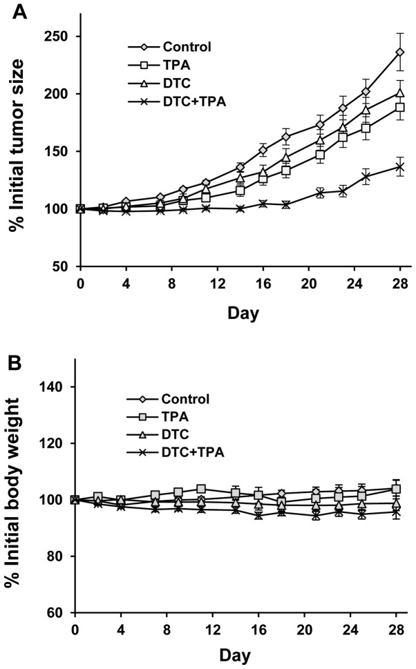
The aim of the present study was to determine the effects of 12-O-tetradecanoylphorbol-13-acetate (TPA) and diethyldithiocarbamate (DDTC) alone or in combination on human pancreatic cancer cells cultured in vitro and grown as xenograft tumors in nude mice. Pancreatic cancer cells were treated with either DDTC or TPA alone, or in combination and the number of viable cells was then determined by trypan blue ecxlusion assay and the number of apoptotic cells was determined by morphological assessment by staining the cells with propidium idiode and examining them under a fluorescence microscope. Treatment with DDTC or TPA alone inhibited the growth and promoted the apoptosis of pancreatic cancer cells in a concentration-dependent manner. These effects were more prominent following treatment with TPA in combination with DDTC than following treatment with either agent alone in PANC-1 cells in monolayer cultures and in 3 dimensional (3D) cultures. The potent effects of the combination treatment on PANC-1 cells were associated with the inhibition of nuclear factor-κB (NF-κB) activation and the decreased expression of Bcl-2 induced by DDTC, as shown by NF-κB-dependent reporter gene expression assay and western blot analysis. Furthermore, treatment of nude mice with DDTC + TPA strongly inhibited the growth of PANC-1 xenograft tumors. The results of the present study indicate that the administration of TPA and DDTC in combination may be an effective strategy for inhibiting the growth of pancreatic cancer.
Figures


Similar articles
-
A binuclear complex constituted by diethyldithiocarbamate and copper(I) functions as a proteasome activity inhibitor in pancreatic cancer cultures and xenografts.Toxicol Appl Pharmacol. 2013 Dec 15;273(3):477-83. doi: 10.1016/j.taap.2013.09.009. Epub 2013 Sep 20. Toxicol Appl Pharmacol. 2013. PMID: 24060341
-
Effects of 12-O-tetradecanoylphorbol-13-acetate in combination with gemcitabine on Panc-1 pancreatic cancer cells cultured in vitro or Panc-1 tumors grown in immunodeficient mice.Int J Oncol. 2012 Dec;41(6):2269-75. doi: 10.3892/ijo.2012.1651. Epub 2012 Oct 4. Int J Oncol. 2012. PMID: 23041978
-
Combination of diethyldithiocarbamate with 12-O-tetradecanoyl phorbol-13-acetate inhibits the growth of human myeloid leukemia HL-60 cells in vitro and in xenograft model.Biosci Biotechnol Biochem. 2020 Oct;84(10):2069-2076. doi: 10.1080/09168451.2020.1789837. Epub 2020 Jul 8. Biosci Biotechnol Biochem. 2020. PMID: 32640883
-
Effects of 12-O-tetradecanoylphorbol-13-acetate (TPA) in combination with paclitaxel (Taxol) on prostate Cancer LNCaP cells cultured in vitro or grown as xenograft tumors in immunodeficient mice.Clin Cancer Res. 2006 Jun 1;12(11 Pt 1):3444-51. doi: 10.1158/1078-0432.CCR-05-2823. Clin Cancer Res. 2006. PMID: 16740769
-
Inhibitory effects of 12-O-tetradecanoylphorbol-13-acetate alone or in combination with all-trans retinoic acid on the growth of cultured human pancreas cancer cells and pancreas tumor xenografts in immunodeficient mice.J Pharmacol Exp Ther. 2005 Oct;315(1):170-87. doi: 10.1124/jpet.105.087585. Epub 2005 Jun 23. J Pharmacol Exp Ther. 2005. PMID: 15976015
Cited by
-
Natural Products as Adjunctive Treatment for Pancreatic Cancer: Recent Trends and Advancements.Biomed Res Int. 2017;2017:8412508. doi: 10.1155/2017/8412508. Epub 2017 Jan 23. Biomed Res Int. 2017. PMID: 28232946 Free PMC article. Review.
-
The Effects and Mechanism of YK-4-279 in Combination with Docetaxel on Prostate Cancer.Int J Med Sci. 2017 Apr 7;14(4):356-366. doi: 10.7150/ijms.18382. eCollection 2017. Int J Med Sci. 2017. PMID: 28553168 Free PMC article.
References
-
- Cao H, Le D, Yang LX. Current status in chemotherapy for advanced pancreatic adenocarcinoma. Anticancer Res. 2013;33:1785–1791. - PubMed
Publication types
MeSH terms
Substances
Grants and funding
LinkOut - more resources
Full Text Sources
Other Literature Sources
Medical
