Review
. 2015 Jun;37(6):687-700.
doi: 10.1002/bies.201400188. Epub 2015 Apr 3.

What is the function of mitochondrial networks? A theoretical assessment of hypotheses and proposal for future research

Affiliations
Review

What is the function of mitochondrial networks? A theoretical assessment of hypotheses and proposal for future research

Hanne Hoitzing et al. Bioessays. 2015 Jun.

Abstract

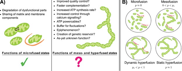
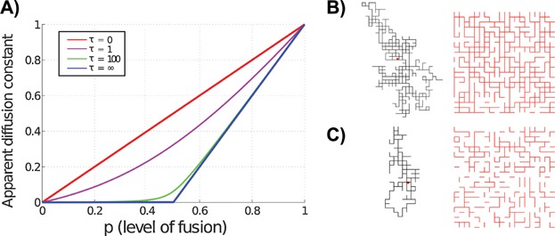
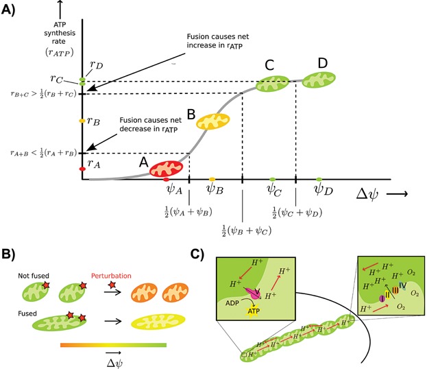
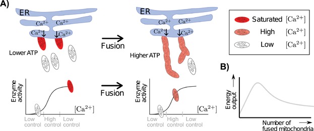
Mitochondria can change their shape from discrete isolated organelles to a large continuous reticulum. The cellular advantages underlying these fused networks are still incompletely understood. In this paper, we describe and compare hypotheses regarding the function of mitochondrial networks. We use mathematical and physical tools both to investigate existing hypotheses and to generate new ones, and we suggest experimental and modelling strategies. Among the novel insights we underline from this work are the possibilities that (i) selective mitophagy is not required for quality control because selective fusion is sufficient; (ii) increased connectivity may have non-linear effects on the diffusion rate of proteins; and (iii) fused networks can act to dampen biochemical fluctuations. We hope to convey to the reader that quantitative approaches can drive advances in the understanding of the physiological advantage of these morphological changes.

Keywords: hypotheses; mathematical biology; mitochondrial dynamics; mitochondrial networks; non-linearities; ultrastructure.


Figures

Figure 1
Figure 1
Potential causes and types of mitochondrial network formation. A: There are clear benefits of small-scale fusion events (microfusion) such as membrane and matrix protein complementation (and possibly mtDNA complementation) and selective degradation. However, to appreciate these advantages, one does not require the formation of large extended networks, the functions of which remains to be elucidated. In this paper we discuss the functions proposed in the right panel. B: The quantity p, defined as λfus/(λfus + λfis), roughly estimates the probability that any two neighbouring mitochondrial units are fused. Note that different rates of fusion and fission can lead to the same connectivity of the network, as long as the ratio λfus/(λfus + λfis) remains constant. In a static hyperfused state, virtually no fission events occur and therefore no quality control is possible. However, in a dynamic hyperfused state the fission rate is non-zero (λfis > 0) and quality control is present. The red arrows represent fusion or fission events and are absent in the static hyperfused state.
Figure 2
Figure 2
Apparent diffusion coefficient depends non-linearly on degree of fusion. An abrupt change in diffusion rate can occur with only a small change in fusion rate. A: This figure shows the diffusion constant of a particle diffusing on a 2D fluctuating lattice as a function of p (the fraction of present bonds) and τ (the relaxation time of the fluctuating bonds). If τ = ∞, the bonds are static; conversely, τ = 0 corresponds to the limit of very fast fluctuating bonds. B: A single trajectory of a diffusing particle (on the left) and a lattice snapshot for p = 0.6 and τ = 100 (on the right). The red dot in the trajectory marks the starting point of the particle. Existing bonds in the lattice snapshot are shown in red. C: Trajectory and lattice snapshot for p = 0.4 and τ = 100. Figure B and C show that (for τ = 100) increasing the value of p from 0.4 to 0.6, results in a more connected network and less restricted diffusion, as is also suggested in Fig. A which indicates a rather abrupt increase in effective diffusion rate around p = 0.5.
Figure 3
Figure 3
A: The effect of fusion on rate of ATP synthesis depends on the magnitude of the potentials of the pre-fusing mitochondria. In this figure, ri denotes the ATP synthesis rate (rATP) for mitochondrion i and r(i + j) denotes the rate for the fused product of mitochondria i and j. For simplicity we assume equal mitochondrial size; our results still hold when this assumption is relaxed. Because of the non-linear dependence of rATP on Δψ, if two mitochondria in the exponential regime fuse (i.e. mitochondria A and B), then averaging their potentials upon fusion causes the net ATP synthesis rate to decrease. This is because 2 r(A + B) < rA + rB (the fused mitochondrion is twice as large as the pre-fused mitochondria and we thus effectively have two mitochondria post-fusion, each with rate r(A + B)). In the plateau region, rATP does not depend on Δψ, so there will most likely be no Δψ induced change in rATP if two mitochondria in this regime fuse (e.g. mitochondria C and D). If a mitochondrion from the exponential regime fuses with one from the plateau (i.e. mitochondria B and C), net rATP increases because 2 r(B + C) > rB + rC. B: Mitochondrial fusion buffers fluctuations in membrane potential. Opening of the mitochondrial permeability transition pore or changes in ion leakage can lead to depolarizations of mitochondria. These perturbations to the membrane potential will have less effect when mitochondria are fused because their bigger size makes them more robust. C: Mechanism of mitochondrial power cabling. Oxygen concentrations are higher at the periphery of the cell and mitochondria positioned here will pump protons out of their matrix. In the core of the cell, if ATP is required, the ATP synthase will pump protons into the matrix. A proton gradient establishes itself along the mitochondrial cable and protons diffuse thereby transmitting chemical potential. This is the main idea of mitochondrial power cabling; a replacement of diffusion of ATP or oxygen through the cytoplasm by proton movement along mitochondrial filaments, which may result in an increased speed of energy transmission.
Figure 4
Figure 4
Fusion may increase controllability of TCA cycle activity and total energy production. A: If only mitochondria in contact with the ER easily take up calcium, then in a fragmented state only few mitochondria will have high (saturated) [Ca2+]matrix and most will have low [Ca2+]matrix. The bottom figure indicates the position of the mitochondria on the sigmoid relating calcium concentration to enzymatic activity. If mitochondria fuse, [Ca2+]matrix averages out, which moves the previously saturated mitochondria down the sigmoid and also moves the mitochondria which previously had very low [Ca2+]matrix up the sigmoid with the result of increasing total enzyme stimulation. When even more mitochondria fuse, total enzyme stimulation will drop again because the mitochondria moved too far down the sigmoid. B: As the number of mitochondria fused to a mitochondrion close to the ER increases, total energy output will first increase and then decrease again because [Ca2+]matrix is now so diluted as to reach the low enzyme activity regime. This plot is a schematic illustration of principle, details are given in section S1.5.

References

    1. Braschi E, McBride HM. Mitochondria and the culture of the Borg. BioEssays. 2010;32:958–66. - PMC - PubMed
    1. Laar VSV, Berman SB. Mitochondrial dynamics in Parkinson’s disease. Exp Neurol. 2009;218:247–56. - PMC - PubMed
    1. Liesa M, Palacín M, Zorzano A. Mitochondrial dynamics in mammalian health and disease. Physiol Rev. 2009;89:799–845. - PubMed
    1. Grandemange S, Herzig S, Martinou JC. Mitochondrial dynamics and cancer. Semin Cancer Biol. 2009;19:50–6. - PubMed
    1. Zhu X, Perry G, Smith MA, Wang X. Abnormal mitochondrial dynamics in the pathogenesis of Alzheimer’s disease. J Alzheimers Dis. 2013;33:S253–62. - PMC - PubMed

Substances