. 2015 Jun;83(6):2475-86.
doi: 10.1128/IAI.02606-14. Epub 2015 Apr 6.

AMA1-deficient Toxoplasma gondii parasites transiently colonize mice and trigger an innate immune response that leads to long-lasting protective immunity

Affiliations

AMA1-deficient Toxoplasma gondii parasites transiently colonize mice and trigger an innate immune response that leads to long-lasting protective immunity

Vanessa Lagal et al. Infect Immun. 2015 Jun.

Abstract

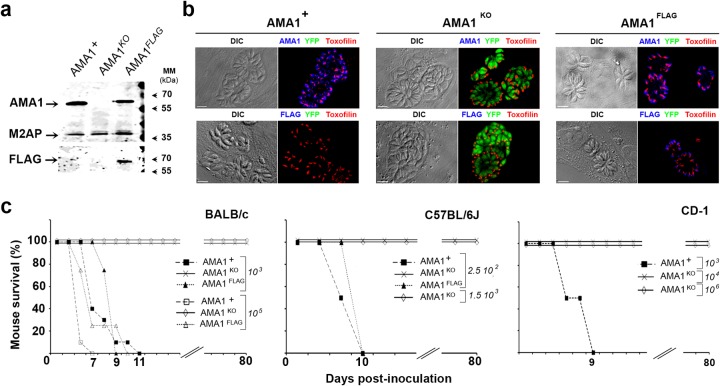
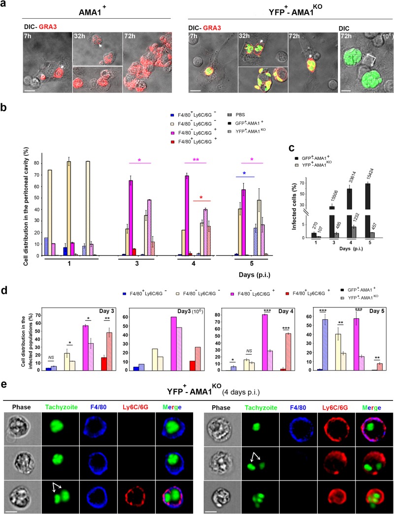
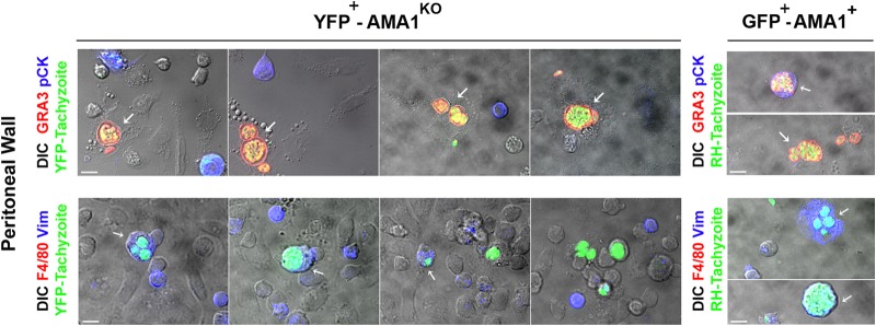
The apical membrane antigen 1 (AMA1) protein was believed to be essential for the perpetuation of two Apicomplexa parasite genera, Plasmodium and Toxoplasma, until we genetically engineered viable parasites lacking AMA1. The reduction in invasiveness of the Toxoplasma gondii RH-AMA1 knockout (RH-AMA1(KO)) tachyzoite population, in vitro, raised key questions about the outcome associated with these tachyzoites once inoculated in the peritoneal cavity of mice. In this study, we used AMNIS technology to simultaneously quantify and image the parasitic process driven by AMA1(KO) tachyzoites. We report their ability to colonize and multiply in mesothelial cells and in both resident and recruited leukocytes. While the RH-AMA1(KO) population amplification is rapidly lethal in immunocompromised mice, it is controlled in immunocompetent hosts, where immune cells in combination sense parasites and secrete proinflammatory cytokines. This innate response further leads to a long-lasting status immunoprotective against a secondary challenge by high inocula of the homologous type I or a distinct type II T. gondii genotypes. While AMA1 is definitively not an essential protein for tachyzoite entry and multiplication in host cells, it clearly assists the expansion of parasite population in vivo.


Figures

FIG 1
FIG 1
Reduced virulence of the AMA1KO parasites in mice. (a and b) AMA1 protein expression detected in whole-cell extracts of AMA1+, AMA1KO, and AMA1FLAG parasites using anti-AMA1 (top panel) and anti-FLAG (bottom panel) antibodies (M2AP micronemal protein served as a loading control) (a) and in tachyzoites in host cells (differential inference contrast [DIC] [left] and fluorescent [right] images are shown [AMA1 and AMA1FLAG, blue; toxofilin protein, red]) (b). MM, molecular mass. (c) Mouse survival curves for BALB/c, C57BL/6J, and CD-1 mice injected i.p. with different doses of AMA1KO or AMA1+ or AMA1FLAG tachyzoites. Bars, 10 µm.
FIG 2
FIG 2
AMA1KO tachyzoites induce a moderate proinflammatory cytokine response. (a) IL-6, CCL2, and IL-12 cytokine concentrations in the peritoneal cavity of mice inoculated with PBS, AMA1KO, AMA1+, and AMA1FLAG at days 1, 3, and 5 p.i. Data represent the results of one experiment representative of three independent assays and are expressed in severalfold values with PBS as a reference. (b) Fluorescence-activated cell sorter (FACS) enumeration of cells collected in the peritoneal exudates at days 1, 3, and 5 p.i. Data represent mean values ± standard errors of the means (SEM) of the results of 1 assay representative of 3.
FIG 3
FIG 3
AMA1KO and AMA1+ tachyzoites colonize resident and inflowing immune cells. (a) Cells from the peritoneal fluids of mice inoculated with 105 DiCre YFP AMA1+ (left panel) or 105 or 106 DiCre YFP+ AMA1KO (right panel). The GRA3 protein was detected in growing parasites and in their surrounding PV/PVM (red; the white arrowheads point to the vacuoles, while white arrows point to the large vacuoles frequently seen with the 106 inoculum). (b to e) AMNIS analysis of cells from peritoneal exudates of mice inoculated with 105 GFP+ AMA1+ or YFP+ AMA1KO and labeled for F4/80 and Ly6C/6G. (b) Relative distributions of cell subpopulations while mutant and wild-type tachyzoites are fluorescent. (c) Percentages of infected cells, with the numbers of cells analyzed indicated on top of each column. (d) Relative distributions between the infected cell subpopulations. (e) Images for each type of peritoneal cell infected by AMA1KO parasites at day 4 p.i. Bars, 10 μM.
FIG 4
FIG 4
Neutrophils are hosting AMA1KO and AMA1+ tachyzoites. (a, b, and f) AMNIS images of cells hosting GFP+-AMA1+ tachyzoites (a and b) or YFP+-AMA1KO tachyzoites (b and f) at days 3 to 5 p.i. (a and b) and days 8, 12, and 15 p.i. (f). In panel a, the white arrows point to growing tachyzoites; in panel b, the white arrows (frames for green fluorescence) and arrowheads (merged frames) indicate the neutrophil (LY6G+) subset hosting tachyzoites; in panel f, a single dying tachyzoite (white arrowhead) in an inflammatory monocyte (white arrow) is engulfed by a macrophage. (c, d, and e) Histograms showing the distributions of F4/80 cell subpopulations hosting AMA1KO and AMA1+ tachyzoites (c), infected cells carrying a single parasite or two or more parasites (d), and total peritoneal AMA1KO-infected cell subpopulations at days 8, 12, and 15 p.i. (the amount of cells analyzed by AMNIS is indicated on top of each column) (e, top panel) and plaque assays on HFF monolayers (e, bottom panel). Histogram shows the average number of days required to fully lyse a HFF monolayer after addition of 105 peritoneal cells collected over time p.i. with AMA1KO tachyzoites. Because no HFF lysis was observed when 105 peritoneal cells collected at day 15 p.i. were deposited (time to lyse infinite), we also incubated the HFF monolayer with 106 cells harvested at that latest time point p.i. The images (right frame) show the area of cell lysis increasing with culture time (days 9 and 12) when 105 peritoneal cells collected at day 12 p.i. were added to HFF monolayers. Bars, 10 μM.
FIG 5
FIG 5
AMA1KO tachyzoites massively colonize mesothelium. DIC-fluorescent merged images of cells released from the mesothelium of mice inoculated with YFP+-AMA1KO (left panels) and GFP+-AMA1+ (right panels) tachyzoites were stained for F4/80 molecules (red, bottom panel) or cytokeratin (blue, top panel) or vimentin (blue, bottom panel) proteins; parasitophorous vacuoles and parasites were stained for GRA3 protein (red, top panel). The white arrows indicated the cells containing tachyzoites. Bars in all panels, 10 μM.
FIG 6
FIG 6
Mice infected with AMA1KO parasites seroconvert and are protected against new infections. (a) (Left panel) Reactivity of sera from AMA1KO-inoculated mice 6 to 8 weeks p.i. Western blots show results from 1 representative mouse for each dose of AMA1KO parasites injected. The inoculation doses are indicated on top of each lane: 0 and + refer to the reactivity of sera from a naive mouse and a mouse infected with the cystogenic type II PruΔKU80 strain, respectively. (Right panel) Histogram showing the rate of seroconversion of mice. The number of mice analyzed is indicated on top of each column. (b) Mouse survival curves after immunization with different doses of AMA1KO tachyzoites and challenges with various amounts of type I virulent AMA1+ tachyzoites. The numbers indicated in the graphs are the inoculum doses at which mice survived. The mice were then challenged with 105 AMA1+ parasites for BALB/c and CD1 mice and 103 AMA1+ parasites for C57BL6 mice. (c) Parasite burden in the brain of infected BALB/c mice challenged (n = 5) or not (n = 2) by AMA1KO parasites and then i.p. injected with 103 type II (ME49) tachyzoites 8 weeks later. A T. gondii RT-PCR assay was performed on brain tissue extracts 6 weeks postinfection, and data are calibrated with known numbers of parasites.

References

    1. Thompson RC, Kutz SJ, Smith A. 2009. Parasite zoonoses and wildlife: emerging issues. Int J Environ Res Public Health 6:678–693. doi:10.3390/ijerph6020678. - DOI - PMC - PubMed
    1. Hill DE, Dubey JP. 2013. Toxoplasma gondii prevalence in farm animals in the United States. Int J Parasitol 43:107–113. doi:10.1016/j.ijpara.2012.09.012. - DOI - PubMed
    1. Pappas G, Roussos N, Falagas ME. 2009. Toxoplasmosis snapshots: global status of Toxoplasma gondii seroprevalence and implications for pregnancy and congenital toxoplasmosis. Int J Parasitol 39:1385–1394. doi:10.1016/j.ijpara.2009.04.003. - DOI - PubMed
    1. Montoya JG, Liesenfeld O. 2004. Toxoplasmosis. Lancet 363:1965–1976. doi:10.1016/S0140-6736(04)16412-X. - DOI - PubMed
    1. Su C, Khan A, Zhou P, Majumdar D, Ajzenberg D, Dardé ML, Zhu XQ, Ajioka JW, Rosenthal BM, Dubey JP, Sibley LD. 2012. Globally diverse Toxoplasma gondii isolates comprise six major clades originating from a small number of distinct ancestral lineages. Proc Natl Acad Sci U S A 109:5844–5849. doi:10.1073/pnas.1203190109. - DOI - PMC - PubMed