

αβ TCR-mediated recognition: relevance to tumor-antigen discovery and cancer immunotherapy
- PMID: 25847967
- PMCID: PMC4391277
- DOI: 10.1158/2326-6066.CIR-15-0042
αβ TCR-mediated recognition: relevance to tumor-antigen discovery and cancer immunotherapy
Abstract
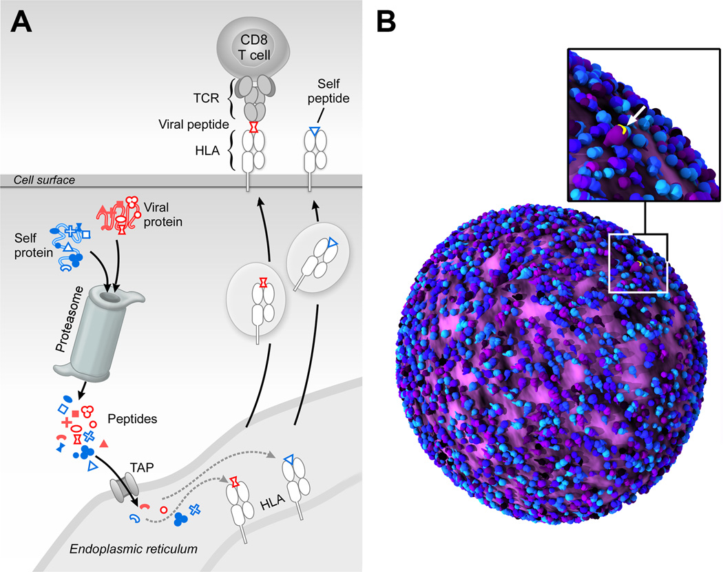
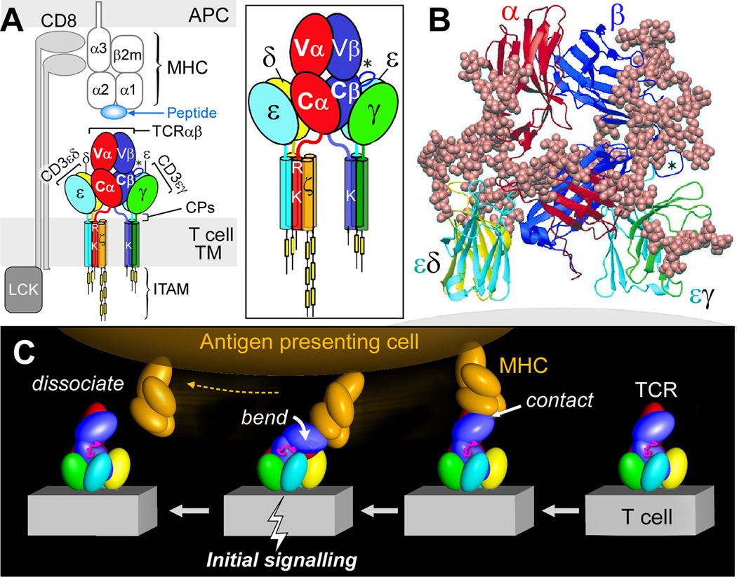
αβ T lymphocytes sense perturbations in host cellular body components induced by infectious pathogens, oncogenic transformation, or chemical or physical damage. Millions to billions of these lymphocytes are generated through T-lineage development in the thymus, each endowed with a clonally restricted surface T-cell receptor (TCR). An individual TCR has the capacity to recognize a distinct "foreign" peptide among the myriad of antigens that the mammalian host must be capable of detecting. TCRs explicitly distinguish foreign from self-peptides bound to major histocompatibility complex (MHC) molecules. This is a daunting challenge, given that the MHC-linked peptidome consists of thousands of distinct peptides with a relevant nonself target antigen often embedded at low number, among orders of magnitude higher frequency self-peptides. In this Masters of Immunology article, I review how TCR structure and attendant mechanobiology involving nonlinear responses affect sensitivity as well as specificity to meet this requirement. Assessment of human tumor-cell display using state-of-the-art mass spectrometry physical detection methods that quantify epitope copy number can help to provide information about requisite T-cell functional avidity affording protection and/or therapeutic immunity. Future rational CD8 cytotoxic T-cell-based vaccines may follow, targeting virally induced cancers, other nonviral immunogenic tumors, and potentially even nonimmunogenic tumors whose peptide display can be purposely altered by MHC-binding drugs to stimulate immune attack.
© 2015 American Association for Cancer Research.
Figures
References
-
- Tonegawa S. In: Nobel Lectures: Physiology or Medicine 1981 – 1990. Frångsmyr T, Lindsten J, editors. Singapore: World Scientific Publishing; 1993. pp. 381–405.
-
- Doherty PC. The nobel lectures in immunology. The Nobel prize for physiology or medicine, 1996; Cell mediated immunity in virus infections. Scand J Immunol. 1996;46:527–540. - PubMed
-
- Zinkernagel RM. The nobel lectures in immunology. The Nobel prize for physiology or medicine, 1996; Cellular immune recognition and the biological role of major transplantation antigens. Scand J Immunol. 1997;46:421–436. - PubMed
Publication types
MeSH terms
Substances
Grants and funding
LinkOut - more resources
Full Text Sources
Research Materials
