

Glucocorticoid receptor regulates accurate chromosome segregation and is associated with malignancy
- PMID: 25847991
- PMCID: PMC4418855
- DOI: 10.1073/pnas.1411356112
Glucocorticoid receptor regulates accurate chromosome segregation and is associated with malignancy
Abstract
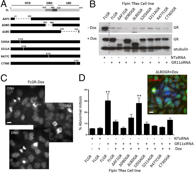
The glucocorticoid receptor (GR) is a member of the nuclear receptor superfamily, which controls programs regulating cell proliferation, differentiation, and apoptosis. We have identified an unexpected role for GR in mitosis. We discovered that specifically modified GR species accumulate at the mitotic spindle during mitosis in a distribution that overlaps with Aurora kinases. We found that Aurora A was required to mediate mitosis-driven GR phosphorylation, but not recruitment of GR to the spindle. GR was necessary for mitotic progression, with increased time to complete mitosis, frequency of mitotic aberrations, and death in mitosis observed following GR knockdown. Complementation studies revealed an essential role for the GR ligand-binding domain, but no clear requirement for ligand binding in regulating chromosome segregation. The GR N-terminal domain, and specifically phosphosites S203 and S211, were not required. Reduced GR expression results in a cell cycle phenotype, with isolated cells from mouse and human subjects showing changes in chromosome content over prolonged passage. Furthermore, GR haploinsufficient mice have an increased incidence of tumor formation, and, strikingly, these tumors are further depleted for GR, implying additional GR loss as a consequence of cell transformation. We identified reduced GR expression in a panel of human liver, lung, prostate, colon, and breast cancers. We therefore reveal an unexpected role for the GR in promoting accurate chromosome segregation during mitosis, which is causally linked to tumorigenesis, making GR an authentic tumor suppressor gene.
Keywords: DNA damage; aneuploidy; cancer; glucocorticoid receptor; mitosis.
Conflict of interest statement
The authors declare no conflict of interest.
Figures


Similar articles
-
Cell cycle phase regulates glucocorticoid receptor function.PLoS One. 2011;6(7):e22289. doi: 10.1371/journal.pone.0022289. Epub 2011 Jul 29. PLoS One. 2011. PMID: 21829454 Free PMC article.
-
USP44 regulates centrosome positioning to prevent aneuploidy and suppress tumorigenesis.J Clin Invest. 2012 Dec;122(12):4362-74. doi: 10.1172/JCI63084. Epub 2012 Nov 26. J Clin Invest. 2012. PMID: 23187126 Free PMC article.
-
The tumor suppressor Lats2 is pivotal in Aurora A and Aurora B signaling during mitosis.Cell Cycle. 2011 Aug 15;10(16):2724-36. doi: 10.4161/cc.10.16.16873. Epub 2011 Aug 15. Cell Cycle. 2011. PMID: 21822051
-
The Aurora kinases: role in cell transformation and tumorigenesis.Cancer Metastasis Rev. 2003 Dec;22(4):451-64. doi: 10.1023/a:1023789416385. Cancer Metastasis Rev. 2003. PMID: 12884918 Review.
-
Mitotic spindle multipolarity without centrosome amplification.Nat Cell Biol. 2014 May;16(5):386-94. doi: 10.1038/ncb2958. Nat Cell Biol. 2014. PMID: 24914434 Review.
Cited by
-
Reciprocal and Autonomous Glucocorticoid and Androgen Receptor Activation in Salivary Duct Carcinoma.Clin Cancer Res. 2020 Mar 1;26(5):1175-1184. doi: 10.1158/1078-0432.CCR-19-1603. Epub 2019 Nov 26. Clin Cancer Res. 2020. PMID: 31772120 Free PMC article.
-
A dual role for glucocorticoid-induced leucine zipper in glucocorticoid function: tumor growth promotion or suppression?Cell Death Dis. 2018 May 1;9(5):463. doi: 10.1038/s41419-018-0558-1. Cell Death Dis. 2018. PMID: 29695779 Free PMC article. Review.
-
Glucocorticoid receptor triggers a reversible drug-tolerant dormancy state with acquired therapeutic vulnerabilities in lung cancer.Nat Commun. 2021 Jul 16;12(1):4360. doi: 10.1038/s41467-021-24537-3. Nat Commun. 2021. PMID: 34272384 Free PMC article.
-
A non-transcriptional role for the glucocorticoid receptor in mediating the cell stress response.Sci Rep. 2017 Sep 21;7(1):12101. doi: 10.1038/s41598-017-09722-z. Sci Rep. 2017. PMID: 28935859 Free PMC article.
-
Daily glucocorticoids promote glioblastoma growth and circadian synchrony to the host.Cancer Cell. 2025 Jan 13;43(1):144-160.e7. doi: 10.1016/j.ccell.2024.11.012. Epub 2024 Dec 12. Cancer Cell. 2025. PMID: 39672168
References
-
- Evans RM. The nuclear receptor superfamily: A rosetta stone for physiology. Mol Endocrinol. 2005;19(6):1429–1438. - PubMed
-
- Rhen T, Cidlowski JA. Antiinflammatory action of glucocorticoids—New mechanisms for old drugs. N Engl J Med. 2005;353(16):1711–1723. - PubMed
-
- Davies TH, Ning YM, Sánchez ER. A new first step in activation of steroid receptors: Hormone-induced switching of FKBP51 and FKBP52 immunophilins. J Biol Chem. 2002;277(7):4597–4600. - PubMed
Publication types
MeSH terms
Substances
Grants and funding
LinkOut - more resources
Full Text Sources
Other Literature Sources
Molecular Biology Databases
