. 2015 Apr 21;112(16):5087-92.
doi: 10.1073/pnas.1424435112. Epub 2015 Apr 6.

Insights into the origins of fish hunting in venomous cone snails from studies of Conus tessulatus

Affiliations

Insights into the origins of fish hunting in venomous cone snails from studies of Conus tessulatus

Joseph W Aman et al. Proc Natl Acad Sci U S A. .

Abstract

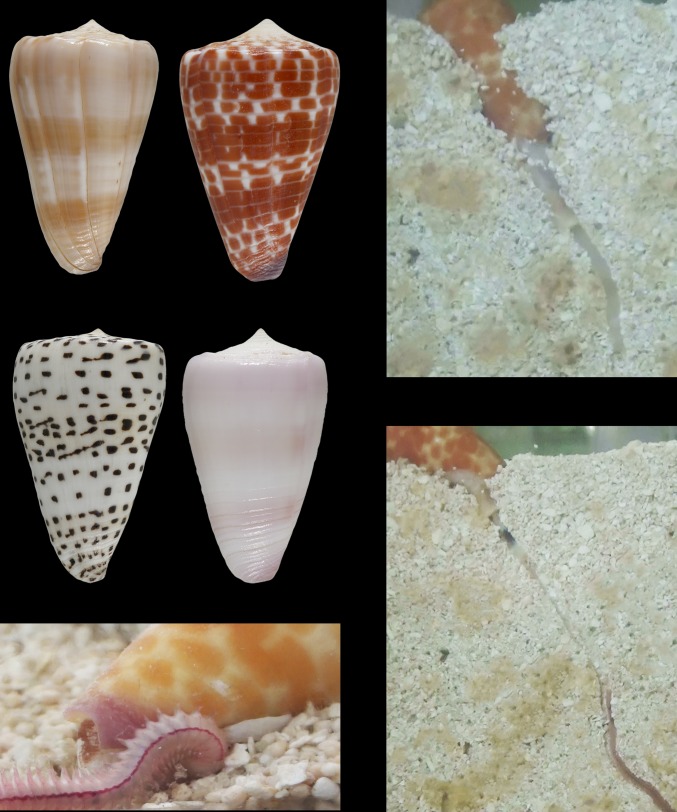
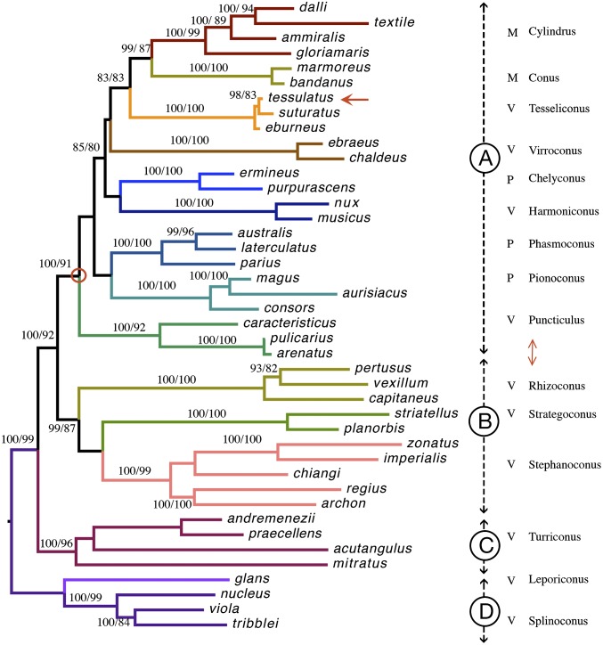
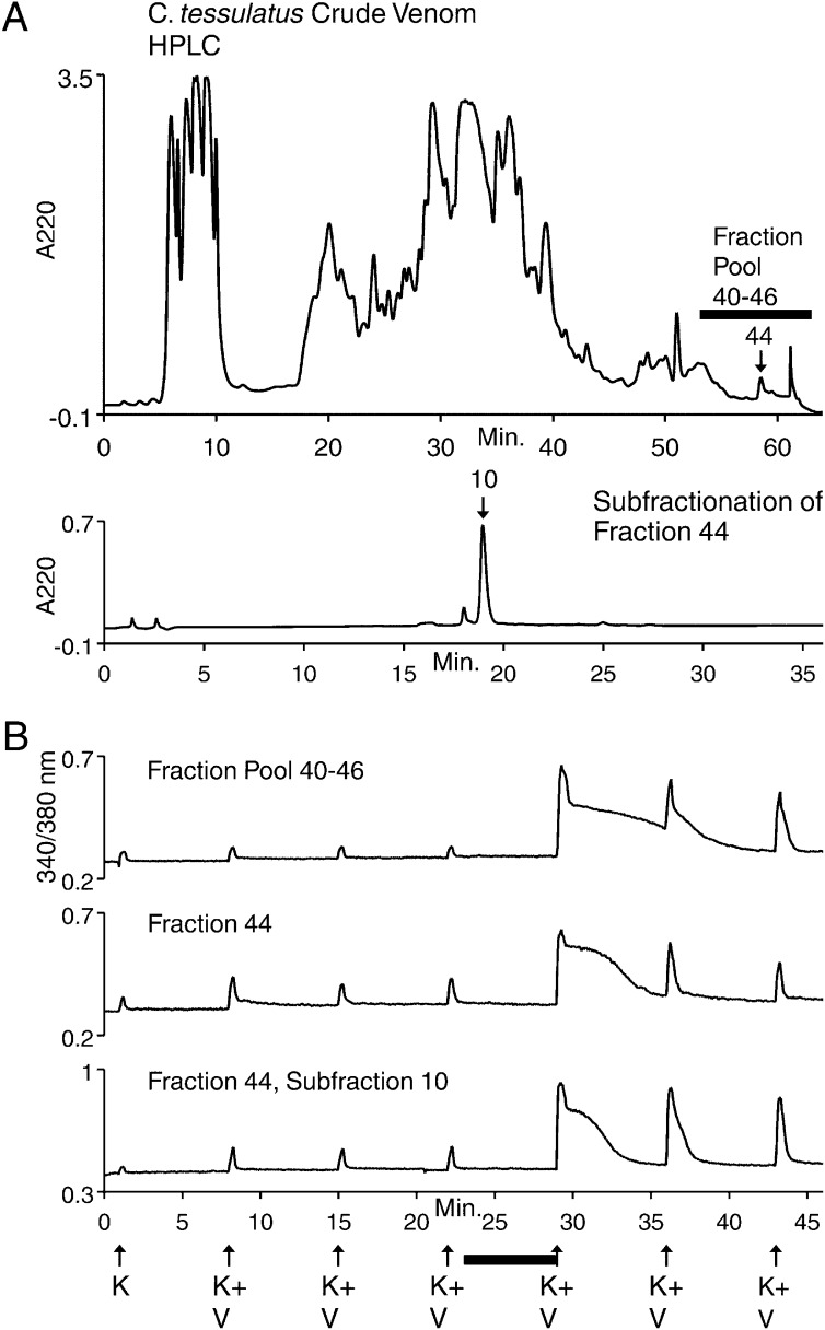
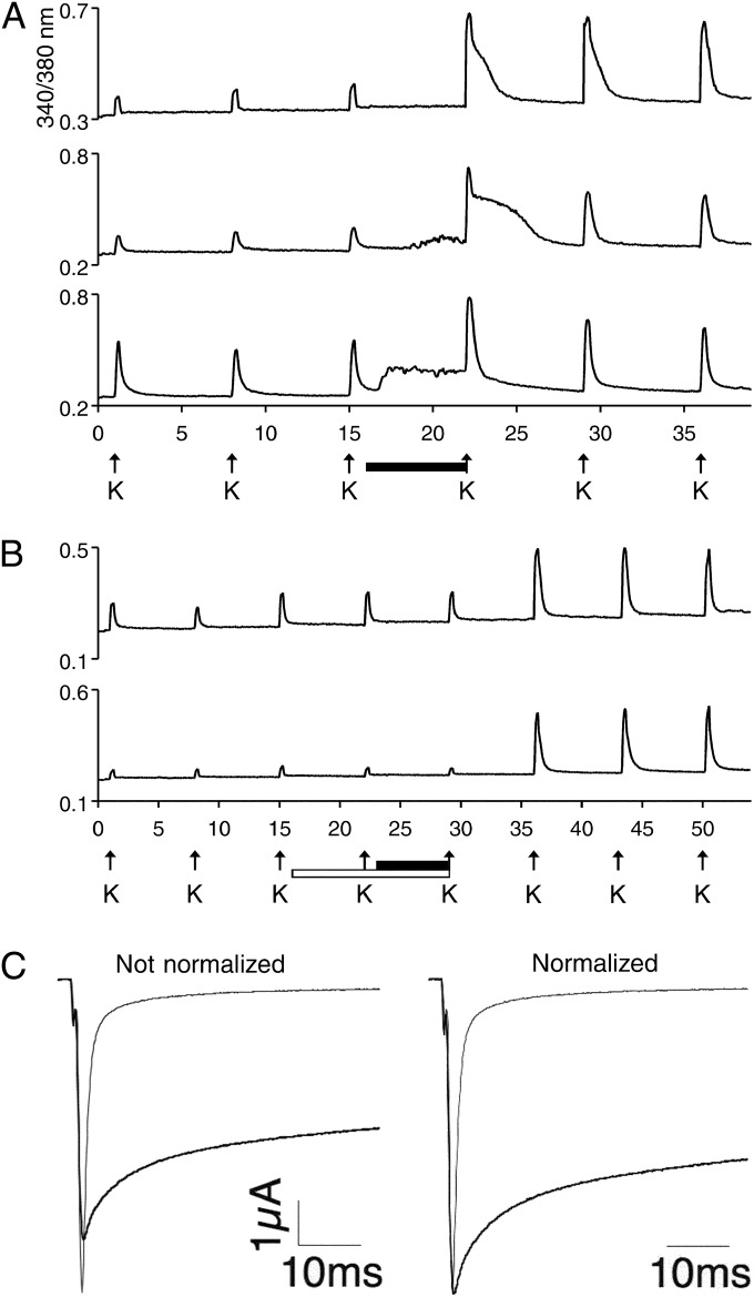
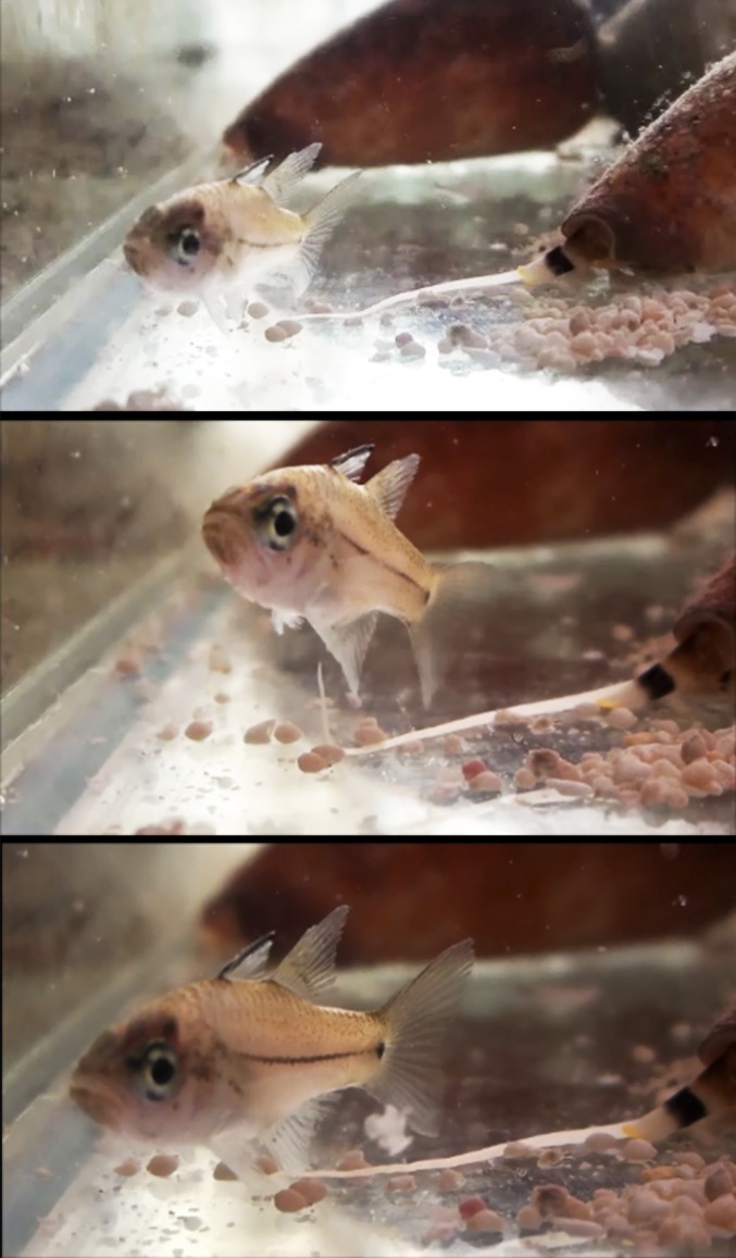
Prey shifts in carnivorous predators are events that can initiate the accelerated generation of new biodiversity. However, it is seldom possible to reconstruct how the change in prey preference occurred. Here we describe an evolutionary "smoking gun" that illuminates the transition from worm hunting to fish hunting among marine cone snails, resulting in the adaptive radiation of fish-hunting lineages comprising ∼100 piscivorous Conus species. This smoking gun is δ-conotoxin TsVIA, a peptide from the venom of Conus tessulatus that delays inactivation of vertebrate voltage-gated sodium channels. C. tessulatus is a species in a worm-hunting clade, which is phylogenetically closely related to the fish-hunting cone snail specialists. The discovery of a δ-conotoxin that potently acts on vertebrate sodium channels in the venom of a worm-hunting cone snail suggests that a closely related ancestral toxin enabled the transition from worm hunting to fish hunting, as δ-conotoxins are highly conserved among fish hunters and critical to their mechanism of prey capture; this peptide, δ-conotoxin TsVIA, has striking sequence similarity to these δ-conotoxins from piscivorous cone snail venoms. Calcium-imaging studies on dissociated dorsal root ganglion (DRG) neurons revealed the peptide's putative molecular target (voltage-gated sodium channels) and mechanism of action (inhibition of channel inactivation). The results were confirmed by electrophysiology. This work demonstrates how elucidating the specific interactions between toxins and receptors from phylogenetically well-defined lineages can uncover molecular mechanisms that underlie significant evolutionary transitions.

Keywords: cone snails; conotoxin; evolution; prey preference.


Conflict of interest statement

The authors declare no conflict of interest.

Figures

Fig. 1.
Fig. 1.
C. tessulatus and related cone snails. (Top Left) Shells of the Tesseliconus clade are shown. Clockwise from top left: C. sandwichensis (Oahu, HI); C. tessulatus (Cocos Island, off Mexico); C. suturatus (Queensland, Australia); and C. eburneus (Cebu, Philippines). C. tessulatus is geographically the most widely distributed of all Conus species. In contrast, C. sandwichensis is an endemic species, restricted to the Hawaiian Islands. (Bottom Left) C. tessulatus engulfing its polychaete worm prey on the surface. (Top Right) Close-up view of C. tessulatus extending its proboscis. (Bottom Right) C. tessulatus attacking worm prey buried under the substrate.
Fig. 2.
Fig. 2.
Phylogeny of C. tessulatus. Maximum-likelihood tree inferred from 12S rRNA, 16S rRNA, and cytochrome oxidase subunit I sequences showing the placement of C. tessulatus (arrow) among other Conus species. Support values on the branches are Bayesian posterior probabilities (Left) expressed as percentages and approximate likelihood ratio statistics (Right). Individual clades (subgenera) are identified by different colors, and the primary prey known for that clade is indicated (M, molluscivorous; P, piscivorous; V, vermivorous). The red circle indicates the divergence between clades that are strictly vermivorous (BD) and a major clade (A) with all of the lineages that use molluscivorous and piscivorous prey-capture strategies.
Fig. 3.
Fig. 3.
Bioassay-guided purification of δ-conotoxin TsVIA from C. tessulatus venom. (A, Top) Reversed-phase HPLC chromatogram of C. tessulatus crude venom. The biological activity was first identified in fraction pool 40–46 and then in fraction 44. (Bottom) Subfractionation of fraction 44 resulted in one main peak (subfraction 10) containing a single peptide that retained the biological activity observed in fraction 44. (B) Calcium-imaging traces from selected DRG neurons. Each trace represents the responses of a single neuron. The experimental protocol shown under the x axis is as follows. Each arrow represents a 15-s application of either 20 mM K+ (K) or 20 mM K+ + 20 μM veratridine (K+V) to depolarize the neurons. Upward deflection of the trace represents an increase in cytosolic calcium concentration. K+V was used in screening venom fractions to facilitate the discovery of antagonists of voltage-gated Na channels. The horizontal bar indicates when the venom fraction was present in the bath solution. Each venom fraction caused a substantial enhancement of the amplitude and duration of the response to a depolarizing stimulus (K+V), which was slowly reversible.
Fig. 4.
Fig. 4.
Biological activity of δ-conotoxin TsVIA. (A and B) Calcium imaging. Each trace represents the responses of a single neuron. Each arrow represents a 15-s application of 20 mM K+ to depolarize the neurons. Upward deflection of the trace represents a transient increase in cytosolic calcium concentration. (A) Calcium-imaging traces from selected DRG neurons in response to purified TsVIA. The horizontal bar indicates when ∼1.6 μM purified TsVIA was present in the bath solution. In different DRG neurons, the peptide caused a substantial enhancement of the response to a depolarizing stimulus (K), with or without a direct increase in the baseline cytosolic calcium concentration. The enhancement of the response to the depolarizing stimulus was slowly reversible following removal of TsVIA. (B) The presence of 1 μM tetrodotoxin in the bath (open horizontal bar), to block voltage-gated Na channels, prevented all effects of TsVIA (closed horizontal bar), until unbound TTX and TsVIA were washed out of the bath. This result suggested that TsVIA acts on TTX-sensitive voltage-gated Na channels and does so with much slower reversibility than TTX. (C) Electrophysiology of voltage-gated sodium channels. The effect of TsVIA on voltage-clamped oocytes coexpressing mouse NaV1.6 with rat NaVβ1 subunits. Shown are Na currents in the absence (thin traces) and presence (thick traces) of TsVIA. Fast inactivation of Na current evident in the control trace was blocked substantially by the presence of TsVIA.
Fig. 5.
Fig. 5.
C. tessulatus attempting to envenomate a fish. The snail has extended its proboscis; shortly after the Middle frame, the tip of the proboscis touched a fin of the fish, and the snail expelled a cloud of venom near the fin, which is visible (Bottom) floating above the extended proboscis and near the lower edge of the tail fin.

References

    1. Michener CD. The Bees of the World. Johns Hopkins Univ Press; Baltimore: 2000.
    1. Jollès J, et al. Episodic evolution in the stomach lysozymes of ruminants. J Mol Evol. 1989;28(6):528–535. - PubMed
    1. Jollès J, et al. Amino acid sequences of stomach and nonstomach lysozymes of ruminants. J Mol Evol. 1990;30(4):370–382. - PubMed
    1. Kornegay JR. Molecular genetics and evolution of stomach and nonstomach lysozymes in the hoatzin. J Mol Evol. 1996;42(6):676–684. - PubMed
    1. Kornegay JR, Schilling JW, Wilson AC. Molecular adaptation of a leaf-eating bird: Stomach lysozyme of the hoatzin. Mol Biol Evol. 1994;11(6):921–928. - PubMed

Publication types

LinkOut - more resources