. 2015 Apr 28;112(17):E2130-8.
doi: 10.1073/pnas.1414242112. Epub 2015 Apr 6.

FtsZ filament capping by MciZ, a developmental regulator of bacterial division

Affiliations

FtsZ filament capping by MciZ, a developmental regulator of bacterial division

Alexandre W Bisson-Filho et al. Proc Natl Acad Sci U S A. .

Abstract

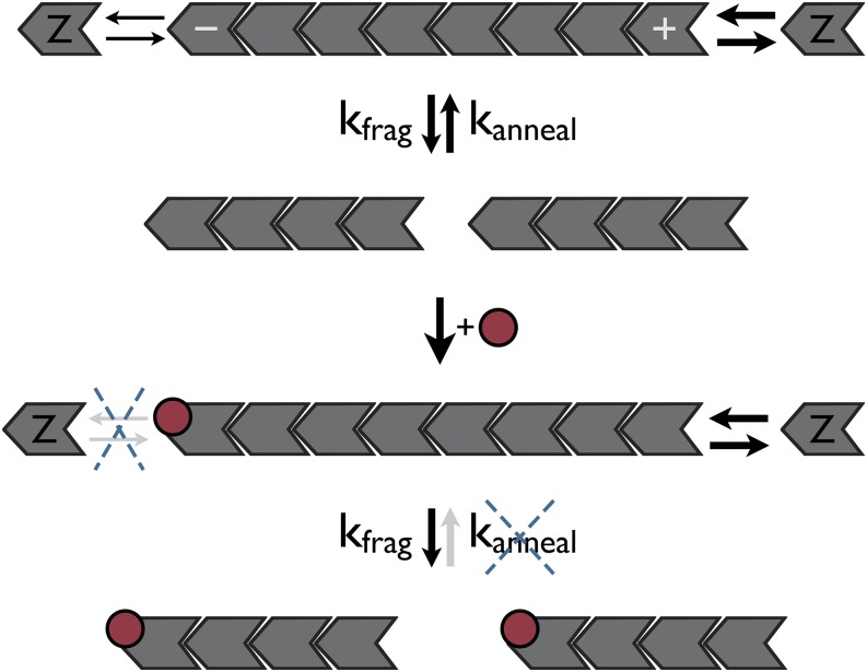
Cytoskeletal structures are dynamically remodeled with the aid of regulatory proteins. FtsZ (filamentation temperature-sensitive Z) is the bacterial homolog of tubulin that polymerizes into rings localized to cell-division sites, and the constriction of these rings drives cytokinesis. Here we investigate the mechanism by which the Bacillus subtilis cell-division inhibitor, MciZ (mother cell inhibitor of FtsZ), blocks assembly of FtsZ. The X-ray crystal structure reveals that MciZ binds to the C-terminal polymerization interface of FtsZ, the equivalent of the minus end of tubulin. Using in vivo and in vitro assays and microscopy, we show that MciZ, at substoichiometric levels to FtsZ, causes shortening of protofilaments and blocks the assembly of higher-order FtsZ structures. The findings demonstrate an unanticipated capping-based regulatory mechanism for FtsZ.

Keywords: FtsZ; bacterial cytoskeleton; cell division; cytokinesis; filament capping.


Conflict of interest statement

The authors declare no conflict of interest.

Figures

Fig. 1.
Fig. 1.
MciZ binds to the minus end of FtsZ and displaces the T7 loop. (A) Cartoon representation of the FtsZ:MciZ complex structure (PDB ID code 4U39). The N-terminal domain of FtsZ (residues 13–178) is represented in blue, the C-terminal domain (residues 209–314) in green, the H7 α-helix (residues 179–202) in yellow, and MciZ (residues 2–37) in red. Cocrystallized phosphates are represented as orange sticks. (B) Superposition of 20 representative low-energy structures of MciZ (red) solved by 1H NMR (PDB ID code 2MRW). Only a segment of the α-helix H1 appears to get structured in absence of FtsZ. (C) Alignment between the FtsZ:MciZ complex (FtsZ in green/blue/yellow and MciZ in red) and the FtsZ monomer of B. subtilis (gray, PDB ID code 2VAM) showing the overlap between MciZ and the T7 loop of the monomer structure, and the absence of the T7 loop in the FtsZ:MciZ structure (blue dotted line). (D) Superposition of MciZ (red, surface rendering) onto the FtsZ dimer structure of S. aureus (PDB 3VOA). The B. subtilis FtsZ:MciZ complex structure was aligned with the upper subunit of the dimer (gray FtsZ). Note the steric clash between MciZ and the bottom subunit of the dimer (orange FtsZ).
Fig. 2.
Fig. 2.
MciZ inhibits FtsZ polymerization substoichiometrically in vitro. All assembly experiments were done in HMK buffer (50 mM Hepes, 5 mM MgAc, 100 mM KAc, pH 7.7). (A and B) Effect of various concentrations (0, 0.2, 0.5, 1, and 2.5 µM) of MciZ (A) or the FtsZ-MciZ fusion (B) on the polymerization of FtsZ (5 µM) reported by right-angle light scattering. (C–E) Effect of MciZ or FtsZ-MciZ on FtsZ filament length visualized by negative-staining EM. For this, 3 µM FtsZ was polymerized without (C) and with 0.3 µM MciZ (D) or 0.3 µM FtsZ-MciZ (E). The filament length distribution in each situation was measured and is plotted (Right). (Scale bar, 100 nm.)
Fig. 3.
Fig. 3.
MciZ blocks cell division substoichiometrically in vivo. (A and B) B. subtilis cells without (A) and with (B) GFP-MciZ expression induced by 0.1% xylose for 2 h. The cells were stained with FM 5-95 membrane dye to reveal septa. (Scale bar, 3 µm.) (C) Quantification of GFP-MciZ from cell extracts. GFP-MciZ was induced with 0.1% xylose for 2 h. GFP-MciZ was detected by immunoblotting using anti-GFP antibody and quantification was carried out by comparing the intensities of the bands with that of purified GFP standards.
Fig. 4.
Fig. 4.
MciZ binds to FtsZ filaments. (A and B) Cosedimentation of FtsZ polymers and MciZ. FtsZ was added to the reaction to a final concentration of 10 µM (9 µM of unlabeled wild type FtsZ and 1 µM of FtsZ-S152C labeled with FITC). MciZ-W36C and MciZ-R20D+W36C were labeled with TMR and added to a final concentration of 250 nM (when indicated). Fluorescent measurements from supernatant (light gray) and pellet (dark gray) fractions were obtained by excitation at 495 nm (FITC) and 550 nm (TMR). NF, normalized fluorescence; P, pellet; S, supernatant. (C) Colocalization of Ftsz-mCherry and GFP-MciZ in B. subtilis live cells. (Upper) Cells expressing FtsZ-mCherry (Left) and GFP-MciZ (Right). (Lower) The mislocalization of GFP-MciZ(R20D) mutant compared with the wild-type MciZ. (Scale bar, 2 µm.)
Fig. 5.
Fig. 5.
MciZ enhances the turnover of FtsZ filaments. (A) FtsZ polymerization reported by fluorescence quenching with BODIPY-conjugated FtsZ-S152C+S223W. Assembly curves with different FtsZ and MciZ concentrations were run to steady state and the ΔFluorescence was plotted. MciZ concentrations are indicated above the lines. (B) Effect of MciZ on the GTPase activity of FtsZ. (C and D) Kinetics of disassembly of FtsZ protofilaments reported by BODIPY-quench system: (C) in 5 mM Mg2+ (HMK buffer, which supports GTPase on), (D) in 1 mM EDTA (MEK buffer, GTPase blocked). Five micromolars of FtsZ was preassembled in the absence (gray) or presence of 0.5 µM of MciZ (red) and the rate of disassembly was measured after dilution in the appropriate buffer.
Fig. 6.
Fig. 6.
Mechanism of FtsZ polymerization inhibition by MciZ. FtsZ filament length is determined by the rates of subunit gain and loss at filament ends, and the balance between filament fragmentation (kfrag) and annealing (kanneal). Capping of filament minus ends by MciZ (red circles) will inhibit subunit addition at these ends, but in principle, growth at the free plus ends could compensate that. In contrast, inhibition of annealing by MciZ turns fragmentation into an irreversible reaction, leading to shortening of FtsZ filaments. At close to stoichiometric concentrations, MciZ should also promote subunit sequestration in addition to capping.

Similar articles

Cited by

References

    1. Theriot JA. Why are bacteria different from eukaryotes? BMC Biol. 2013;11:119. - PMC - PubMed
    1. Bi EF, Lutkenhaus J. FtsZ ring structure associated with division in Escherichia coli. Nature. 1991;354(6349):161–164. - PubMed
    1. Nogales E, Downing KH, Amos LA, Löwe J. Tubulin and FtsZ form a distinct family of GTPases. Nat Struct Biol. 1998;5(6):451–458. - PubMed
    1. Adams DW, Errington J. Bacterial cell division: Assembly, maintenance and disassembly of the Z ring. Nat Rev Microbiol. 2009;7(9):642–653. - PubMed
    1. Erickson HP, Anderson DE, Osawa M. FtsZ in bacterial cytokinesis: Cytoskeleton and force generator all in one. Microbiol Mol Biol Rev. 2010;74(4):504–528. - PMC - PubMed

Publication types

MeSH terms

LinkOut - more resources