. 2015 Apr 21;112(16):5075-80.
doi: 10.1073/pnas.1421949112. Epub 2015 Apr 6.

Sall4-Gli3 system in early limb progenitors is essential for the development of limb skeletal elements

Affiliations

Sall4-Gli3 system in early limb progenitors is essential for the development of limb skeletal elements

Ryutaro Akiyama et al. Proc Natl Acad Sci U S A. .

Abstract

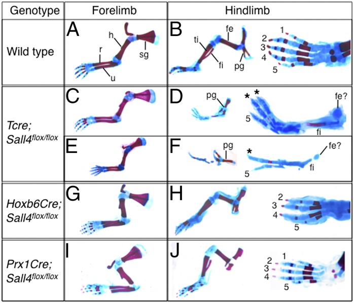
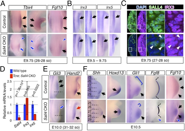
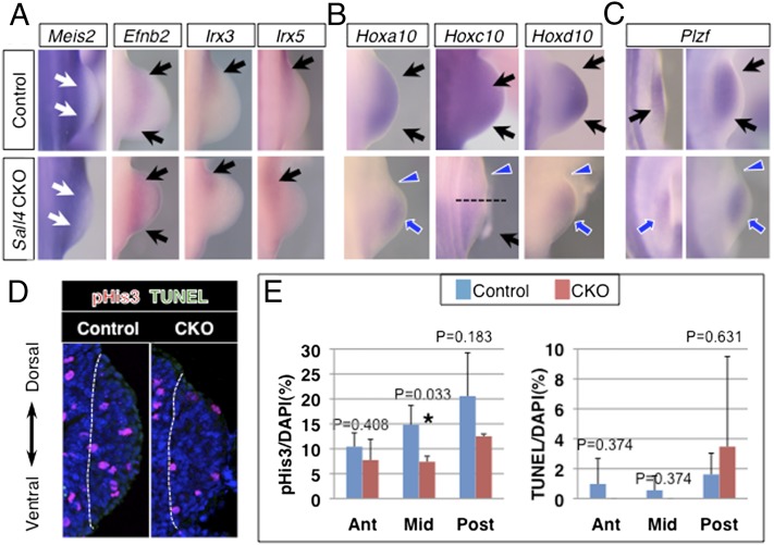
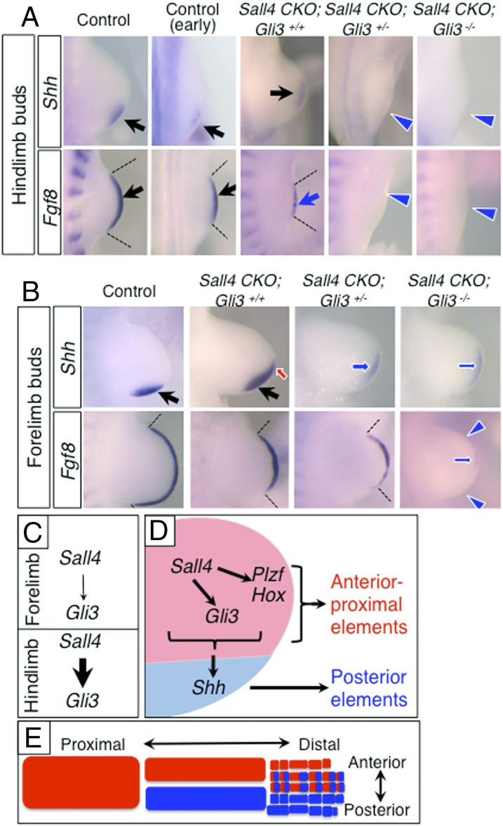
Limb skeletal elements originate from the limb progenitor cells, which undergo expansion and patterning to develop each skeletal element. Posterior-distal skeletal elements, such as the ulna/fibula and posterior digits develop in a Sonic hedgehog (Shh)-dependent manner. However, it is poorly understood how anterior-proximal elements, such as the humerus/femur, the radius/tibia and the anterior digits, are developed. Here we show that the zinc finger factors Sall4 and Gli3 cooperate for proper development of the anterior-proximal skeletal elements and also function upstream of Shh-dependent posterior skeletal element development. Conditional inactivation of Sall4 in the mesoderm before limb outgrowth caused severe defects in the anterior-proximal skeletal elements in the hindlimb. We found that Gli3 expression is reduced in Sall4 mutant hindlimbs, but not in forelimbs. This reduction caused posteriorization of nascent hindlimb buds, which is correlated with a loss of anterior digits. In proximal development, Sall4 integrates Gli3 and the Plzf-Hox system, in addition to proliferative expansion of cells in the mesenchymal core of nascent hindlimb buds. Whereas forelimbs developed normally in Sall4 mutants, further genetic analysis identified that the Sall4-Gli3 system is a common regulator of the early limb progenitor cells in both forelimbs and hindlimbs. The Sall4-Gli3 system also functions upstream of the Shh-expressing ZPA and the Fgf8-expressing AER in fore- and hindlimbs. Therefore, our study identified a critical role of the Sall4-Gli3 system at the early steps of limb development for proper development of the appendicular skeletal elements.

Keywords: Gli3; Plzf-Hox; Sall4; appendicular skeletal elements; limb progenitors.


Conflict of interest statement

The authors declare no conflict of interest.

Figures

Fig. 1.
Fig. 1.
Sall4 is required for development of the anterior-proximal skeletal elements in a temporally restricted manner. (AJ) Appendicular skeletal preparations of control (A and B) and Tcre; Sall4 CKO (CF), Hoxb6Cre; Sall4 CKO (G and H) and Prx1Cre; Sall4 CKO (I and J) neonatal mice. (A, C, E, G, and I) Forelimbs develop without significant defects in mutants with size reduction in severely affected Tcre; Sall4 mutants. (D and F) In Tcre; Sall4 hindlimbs a small cartilage aggregate was present instead of the femur (fe). The fibula (fi) was present, but the tibia (ti) was missing. The pelvic girdle (pg) was small, but patterned normally. (H) In Hoxb6Cre; Sall4 CKO hindlimbs, d1 was missing in the autopod. (J) Hindlimbs of Prx1Cre; Sall4 CKO developed normally. fe, femur; fi, fibula; h, humerus; pg, pelvic girdle; r, radius; ti, tibia; u, ulna. Digits are numbered as 1–5. Asterisks mark digits whose identity is not completely determined.
Fig. 2.
Fig. 2.
Loss of Sall4 caused defects of gene expression in hindlimb progenitors independent of Irx3/5. In situ hybridization (A, B, E, and F), immunofluorescence (C), and quantitative RT-PCR (D) of marker genes/proteins in control and Sall4 CKO hindlimb buds. (A) Dorsal views of Tbx4 and Fgf10, and lateral views of Tbx4 at E9.75. (B) Dorsal views of Irx3 and Irx5 at E9.5-E9.75. (C) Immunofluorescence of SALL4 (green) and IRX3 (magenta) on transverse sections of E9.75 anterior hindlimb buds. White arrowheads point to cells that escaped from Sall4 deletion in Tcre; Sall4 CKO hindlimb buds. (Right) Close up of the boxed areas in Left. (D) Quantitative analysis of Irx3 and Irx5 transcript levels. Relative mRNA levels of indicated genes are shown as average ± SD. Asterisks indicate statistical significance. P values for expression of each gene are included. (E) Dorsal views of Gli3 and Hand2 at E10.0. (F) Dorsal views of Shh, Hoxd13, Gli1, Fgf8 and Fgf10 at E10.5. (A, B, E, and F) Black arrows and blue arrows point to normal and reduced expression, respectively. Red arrows point to ectopic expression. Blue arrowheads denote loss of expression. In D, broken lines mark normal expression border of Gli3. In E, dotted lines mark somite boundaries in Shh expression panels. Brackets in Gli1 expression panels denote anterior domains that are free of Gli1 signals.
Fig. 3.
Fig. 3.
Sall4 integrates Plzf-Hox10 system and proliferative expansion of limb progenitors. (A) Expression pattern of Meis2, Efnb2, Irx3, and Irx5 in control and Sall4 CKO hindlimbs at E10.5. (B) Expression pattern of Hoxa10, Hoxc10, and Hoxd10 in control and Sall4 CKO hindlimbs at E10.5. (C) Expression pattern of Plzf in control and Sall4 CKO hindlimbs at E9.75 and E10.5. Black arrows and blue arrows point to normal and reduced expression, respectively. In A, Meis2 expression is indicated by white arrows. Blue arrowheads denote loss of expression. In B, a broken line indicates the anterior boundary of Hoxc10 expression in Sall4 CKO hindlimb buds. (D) Representative images of pHis3 and TUNEL double staining of transverse sections of nascent hindlimb bud at E9.75 in control and Sall4 CKO embryos. Dotted lines indicate boundary between the nascent hindlimb bud and trunk. (E) Quantitative evaluation of cell proliferation and cell death in transverse sections of nascent hindlimb buds at E9.75 in control and Sall4 CKO embryos. P values are included.
Fig. 4.
Fig. 4.
Genetic interaction between Sall4 and Gli3 and a model of the role of Sall4-Gli3 system in limb progenitor cells. (A) Expression pattern of Shh and Fgf8 in hindlimbs of indicated genotypes. Due to reduced size of hindlimb buds in Sall4 CKO; Gli3 mutants, wild-type hindlimb buds at an earlier stage with similar size were included. (B) Expression pattern of Fgf8 and Shh in forelimbs of indicated genotypes. Black arrows and blue arrows point to normal and reduced expression, respectively. Red arrows point to ectopic expression. Blue arrowheads denote loss of expression. (CE) A model of the role of Sall4 in the two-population model for the development of limb skeletal elements. (C) Sall4 regulation of Gli3 differs in fore- and hindlimb progenitor cells. (D) Model of Sall4 regulation of gene network in the putative anterior progenitors (red) and posterior progenitors (blue). (E) Schematic drawing of skeletal elements derived from the putative anterior progenitors (red) and posterior progenitors (blue).

References

    1. Gros J, Tabin CJ. Vertebrate limb bud formation is initiated by localized epithelial-to-mesenchymal transition. Science. 2014;343(6176):1253–1256. - PMC - PubMed
    1. Zeller R, López-Ríos J, Zuniga A. Vertebrate limb bud development: Moving towards integrative analysis of organogenesis. Nat Rev Genet. 2009;10(12):845–858. - PubMed
    1. Harfe BD, et al. Evidence for an expansion-based temporal Shh gradient in specifying vertebrate digit identities. Cell. 2004;118(4):517–528. - PubMed
    1. Ahn S, Joyner AL. Dynamic changes in the response of cells to positive hedgehog signaling during mouse limb patterning. Cell. 2004;118(4):505–516. - PubMed
    1. Kraus P, Fraidenraich D, Loomis CA. Some distal limb structures develop in mice lacking Sonic hedgehog signaling. Mech Dev. 2001;100(1):45–58. - PubMed

Publication types

MeSH terms

LinkOut - more resources