Review
. 2015 Jun;4(6):555-63.
doi: 10.5966/sctm.2014-0272. Epub 2015 Apr 6.

Concise Review: Macrophages: Versatile Gatekeepers During Pancreatic β-Cell Development, Injury, and Regeneration

Affiliations
Review

Concise Review: Macrophages: Versatile Gatekeepers During Pancreatic β-Cell Development, Injury, and Regeneration

Naomi Van Gassen et al. Stem Cells Transl Med. 2015 Jun.

Abstract

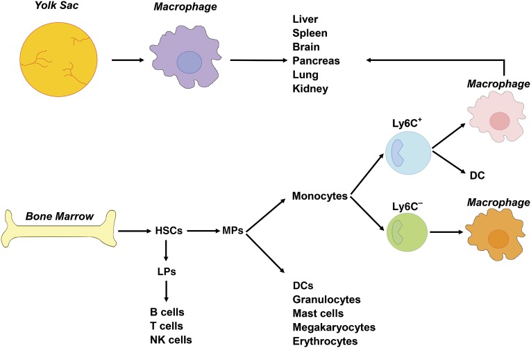
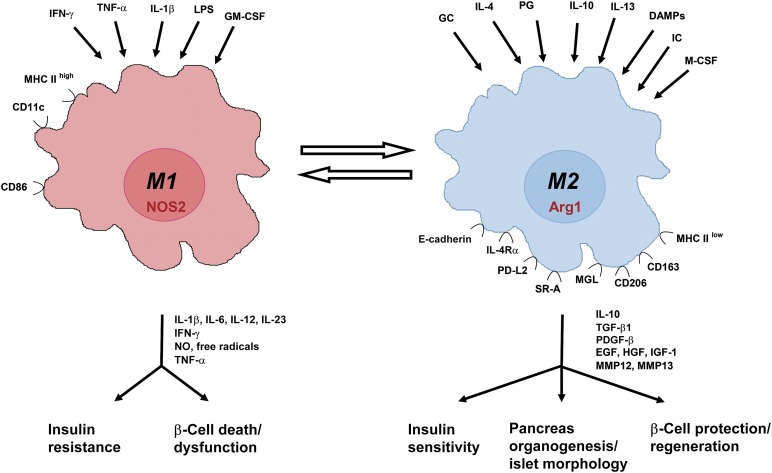
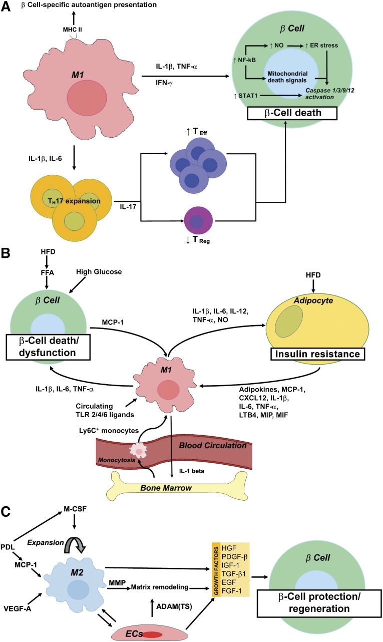
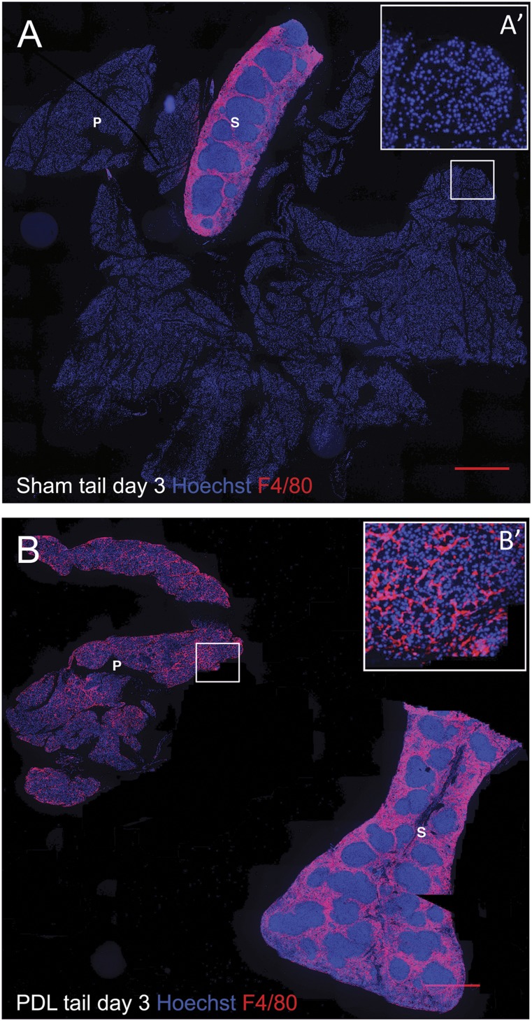
Macrophages are classically considered detrimental for pancreatic β-cell survival and function, thereby contributing to β-cell failure in both type 1 (T1D) and 2 (T2D) diabetes mellitus. In addition, adipose tissue macrophages negatively influence peripheral insulin signaling and promote obesity-induced insulin resistance in T2D. In contrast, recent data unexpectedly uncovered that macrophages are not only able to protect β cells during pancreatitis but also to orchestrate β-cell proliferation and regeneration after β-cell injury. Moreover, by altering their activation state, macrophages are able to improve insulin resistance in murine models of T2D. This review will elaborate on current insights in macrophage heterogeneity and on the evolving role of pancreas macrophages during organogenesis, tissue injury, and repair. Additional identification of macrophage subtypes and of their secreted factors might ultimately translate into novel therapeutic strategies for both T1D and T2D.

Significance: Diabetes mellitus is a pandemic disease, characterized by severe acute and chronic complications. Macrophages have long been considered prime suspects in the pathogenesis of both type 1 and 2 diabetes mellitus. In this concise review, current insights in macrophage heterogeneity and on the, as yet, underappreciated role of alternatively activated macrophages in insulin sensing and β-cell development/repair are reported. Further identification of macrophage subtypes and of their secreted factors might ultimately translate into novel therapeutic strategies for diabetes mellitus.

Keywords: Development; Injury; Macrophage; Pancreas; Regeneration; β Cell.


Figures

Figure 1.
Figure 1.
Macrophage origin in the mouse. Classically, macrophages derive from circulating monocytes, in turn descending from bone marrow progenitors. In the bone marrow, HSCs differentiate into LPs or MPs. LPs further differentiate into B cells, T cells, and NK cells. MPs give rise to DCs, granulocytes, mast cells, megakaryocytes, and erythrocytes, or to Ly6C+ inflammatory and Ly6C resident or patrolling monocytes. After tissue infiltration, Ly6C+ monocytes give rise to either DCs or macrophages. Although less documented, Ly6C monocytes can also differentiate into macrophages. Alternatively, adult tissue macrophages can also descend from yolk sac-derived macrophages, independently of HSCs. As such, tissue macrophages in the adult are mainly a mix of bone marrow-derived and yolk sac-derived macrophages. Abbreviations: DCs, dendritic cells; HSCs, hematopoietic stem cells; LPs, lymphoid committed precursors; MPs, myeloid committed precursors; NK, natural killer (cells).
Figure 2.
Figure 2.
M1 versus M2 macrophages: inducers, markers, effector molecules, and function. Summary of the most important inducers and markers of M1 and M2 macrophages and their role in pancreas development, insulin sensitivity, and β-cell death, dysfunction, and regeneration. Abbreviations: Arg, arginase; CD, cluster of differentiation; DAMPs, damage associated molecular patterns; EGF, epidermal growth factor; GCs, glucocorticoids; GM-CSF, granulocyte-macrophage colony-stimulating factor; HGF, hepatocyte growth factor; ICs, immune complexes; IFN, interferon; IGF, insulin-like growth factor; IL, interleukin; IL-4Rα, IL-4 receptor α; LPS, lipopolysaccharide; M-CSF, macrophage colony-stimulating factor; MGL, macrophage galactose-type lectin; MHC, major histocompatibility complex; MMP, matrix metalloproteinase; NO, nitric oxide; NOS, nitric oxide synthase; PDGF, platelet-derived growth factor; PD-L, programmed death-ligand; PG, prostaglandin; SR, scavenger receptor; TGF, transforming growth factor; TNF, tumor necrosis factor; VEGF, vascular endothelial growth factor.
Figure 3.
Figure 3.
Schematic representation summarizing the role of macrophages in the pathogenesis of type 1 diabetes mellitus (A), type 2 diabetes mellitus (T2D) (B), and during β-cell protection and regeneration (C). (A): M1 macrophages contribute to β-cell death through (a) MHC II-mediated presentation of β-cell-specific autoantigens, (b) IL-1β and IL-6-mediated TH17 expansion and subsequent IL-17-mediated TEff/TReg imbalance, and (c) a direct cytotoxic effect of IL-1β and/or TNF-α plus IFN-γ with downstream activation of NF-κB and STAT1. NF-κB activation results in NO production and increased ER stress and cytochrome c release from mitochondria, the latter acting as a mitochondrial death signal. Ultimately, NF-κB and STAT1 activation results in caspase 1/3/9/12 activation and subsequent β-cell death. (B): High circulating glucose and FFAs contribute to β-cell death in T2D. In addition, a HFD and circulating FFAs promote MCP-1 secretion from β cells and subsequent intraislet accumulation of M1-like macrophages. Moreover, a HFD and onset of T2D correlate with elevated circulating levels of TLR2 and -4 ligands, which stimulate the secretion of proinflammatory cytokines (IL-1β, IL-6, and TNF-α) from these recruited macrophages, thereby further contributing to β-cell death and dysfunction. A high-fat diet and lipid accumulation also promote adipo- (leptin, resistin), chemo-, and cytokine (IL-1β, IL-6, TNF-α, MCP-1, LTB4, CXCL12, MIP, and MIF) secretion from adipocytes, thereby promoting the recruitment to and activation of Ly6C+ monocytes and M1 macrophages in adipose tissue. These exert detrimental effects on peripheral insulin signaling through secretion of IL-1β, IL-6, IL-12, and TNF-α, and production of NO. Finally, M1 adipose tissue macrophages promote myelopoiesis and monocytosis through IL-1β secretion, thereby further enhancing macrophage accumulation in inflamed adipose tissue and, thus, establishing a positive feedback loop in T2D pathogenesis. (C): M2 macrophages accumulate in the pancreas after transgenic, β cell-specific VEGF-A overexpression or partial PDL, in the latter via MCP-1-mediated recruitment and M-CSF-dependent proliferation of macrophages. M2 macrophages promote β-cell protection and regeneration through secretion of several growth factors (i.e., TGF-β1, EGF, PDGF-β, IGF-1) in concert with endothelial cell-derived growth factors (i.e., HGF, FGF-1, IGF-1, TGF-β1, and PDGF-β). M2 macrophages and endothelial cells moreover produce matrix remodeling factors—MMP (i.e., MMP12, MMP13) and ADAM and ADAM(TS) (i.e., ADAM12, ADAMTS9), respectively—facilitating growth factor bioavailability. Abbreviations: ADAM, a disintegrin and metalloproteinase; ADAM(TS), a disintegrin and metalloproteinase with thrombospondin motifs; CXCL, chemokine (C-X-C motif) ligand; EGF, epidermal growth factor; ER, endoplasmic reticulum; FFA, free fatty acid; FGF, fibroblast growth factor; HFD, high-fat diet; IGF, insulin-like growth factor; IL, interleukin; LTB, leukotriene B; MCP, monocyte chemoattractant protein; M-CSF, macrophage colony-stimulating factor; MIF, macrophage migration inhibitory factor; MIP, macrophage inflammatory protein; MMP, matrix metalloproteinase; NF-κB, nuclear factor κB; NO, nitric oxide; PDGF, platelet-derived growth factor; PDL, pancreatic duct ligation; TEff, T effector; TH, T helper; TReg, T regulator; TLR, Toll-like receptor; TNF, tumor necrosis factor; VEGF, vascular endothelial growth factor.
Figure 4.
Figure 4.
Massive macrophage infiltration in the pancreas after partial pancreatic duct ligation. Pancreas tail and spleen sections from day 3 sham-operated (A) and PDL (B) mice, stained for nuclei (Hoechst, blue) and F4/80 (red). (A′, B′): Higher magnification of the area depicted by the squares in (A) and (B). Scale bars = 500 μm. Notably, PDL results in acinar cell loss and thus a decrease in the total pancreas tail area versus sham-operated mice. Abbreviations: P, pancreas tail; PDL, pancreatic duct ligation; S, spleen.

References

    1. Geissmann F, Manz MG, Jung S, et al. Development of monocytes, macrophages, and dendritic cells. Science. 2010;327:656–661. - PMC - PubMed
    1. Wynn TA, Chawla A, Pollard JW. Macrophage biology in development, homeostasis and disease. Nature. 2013;496:445–455. - PMC - PubMed
    1. Schulz C, Gomez Perdiguero E, Chorro L, et al. A lineage of myeloid cells independent of Myb and hematopoietic stem cells. Science. 2012;336:86–90. - PubMed
    1. Ginhoux F, Jung S. Monocytes and macrophages: Developmental pathways and tissue homeostasis. Nat Rev Immunol. 2014;14:392–404. - PubMed
    1. Hashimoto D, Chow A, Noizat C, et al. Tissue-resident macrophages self-maintain locally throughout adult life with minimal contribution from circulating monocytes. Immunity. 2013;38:792–804. - PMC - PubMed

MeSH terms

LinkOut - more resources