

Tissue expanders in reconstruction of maxillofacial defects
- PMID: 25848145
- PMCID: PMC4379297
- DOI: 10.1007/s12663-014-0629-5
Tissue expanders in reconstruction of maxillofacial defects
Abstract
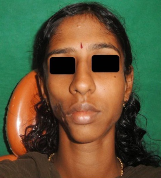
Tissue expansion in its natural ways had fascinated man from prehistoric times itself. But tissue expansion for medical purposes was first tried and reported only in the early half of twentieth century. Presently the principle of tissue expansion is being used in reconstruction of many hard and soft tissue defects of larger dimension, which were previously regarded as great challenge for maxillofacial and plastic surgeons. Making use of the viscoelastic nature of the skin, considerable amount of tissue expansion based tissue engineering is possible in the maxillofacial region. Here we present a case of a facial scar of large dimension with a central oro cutaneous fistula developed as a result of facial artery blow out in a 24 year old female for which esthetic correction was done using the excess tissue obtained from tissue expansion. In this case where other methods of reconstruction such as local flaps, free flaps and normal tissue grafts were assessed to be non viable, tissue expansion was found to be an apt solution for esthetic reconstruction.
Keywords: Fistulous tract; Scar tissue; Tissue expander; Tissue expansion.
Figures


Similar articles
-
Forehead Flap Ballooning for Scar Revision.Ann Maxillofac Surg. 2020 Jan-Jun;10(1):182-185. doi: 10.4103/ams.ams_266_19. Epub 2020 Jun 8. Ann Maxillofac Surg. 2020. PMID: 32855937 Free PMC article.
-
A minimally invasive approach to the placement of tissue expanders.Semin Plast Surg. 2008 Feb;22(1):9-17. doi: 10.1055/s-2007-1019137. Semin Plast Surg. 2008. PMID: 20567683 Free PMC article.
-
The use of tissue expanders in the correction of avulsive injuries of the mandible: report of two cases.J Oral Maxillofac Surg. 1988 Oct;46(10):892-9. doi: 10.1016/0278-2391(88)90059-6. J Oral Maxillofac Surg. 1988. PMID: 3049994
-
Leave the fat, skip the bolster: thinking outside the box in lower third nasal reconstruction.Plast Reconstr Surg. 2004 Nov;114(6):1427-35. doi: 10.1097/01.prs.0000138817.14320.ec. Plast Reconstr Surg. 2004. PMID: 15509929 Review.
-
Tissue engineering technology and its possible applications in oral and maxillofacial surgery.Br J Oral Maxillofac Surg. 2014 Jan;52(1):7-15. doi: 10.1016/j.bjoms.2013.03.005. Epub 2013 Apr 16. Br J Oral Maxillofac Surg. 2014. PMID: 23601833 Review.
Cited by
-
Novel strategies in scalp expansion: improvements and applications of tissue expanders.Burns Trauma. 2024 Apr 8;12:tkae002. doi: 10.1093/burnst/tkae002. eCollection 2024. Burns Trauma. 2024. PMID: 38596624 Free PMC article. No abstract available.
-
Forehead Flap Ballooning for Scar Revision.Ann Maxillofac Surg. 2020 Jan-Jun;10(1):182-185. doi: 10.4103/ams.ams_266_19. Epub 2020 Jun 8. Ann Maxillofac Surg. 2020. PMID: 32855937 Free PMC article.
-
Tissue Expansion Improves the Outcome and Predictability for Alveolar Bone Augmentation: Prospective, Multicenter, Randomized Controlled Trial.J Clin Med. 2020 Apr 16;9(4):1143. doi: 10.3390/jcm9041143. J Clin Med. 2020. PMID: 32316310 Free PMC article.
References
-
- Radovan C (1976) Adjacent flap development using expandable silastic implant. Presented at the annual meeting of the American Society of Plastic and Reconstructive Surgeons, Boston
Publication types
LinkOut - more resources
Full Text Sources
Other Literature Sources