. 2015 Mar 23:10:2325-33.
doi: 10.2147/IJN.S75174. eCollection 2015.

Water-soluble L-cysteine-coated FePt nanoparticles as dual MRI/CT imaging contrast agent for glioma

Affiliations

Water-soluble L-cysteine-coated FePt nanoparticles as dual MRI/CT imaging contrast agent for glioma

Shuyan Liang et al. Int J Nanomedicine. .

Abstract

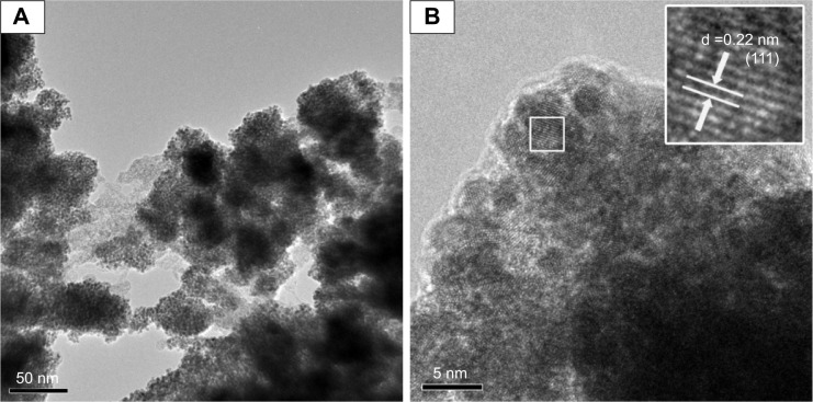
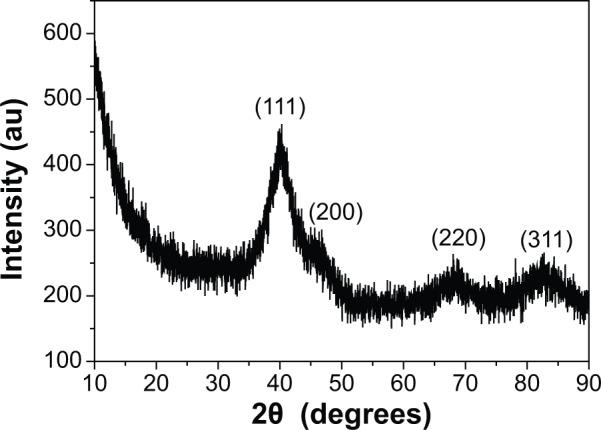
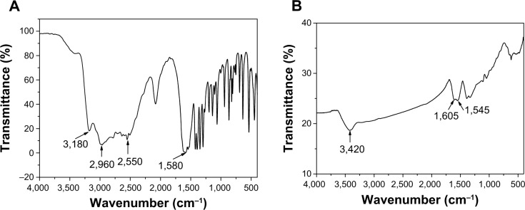
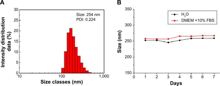
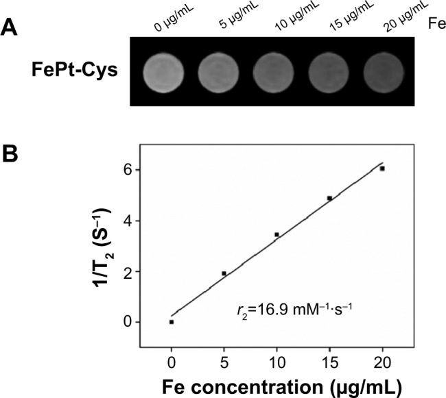
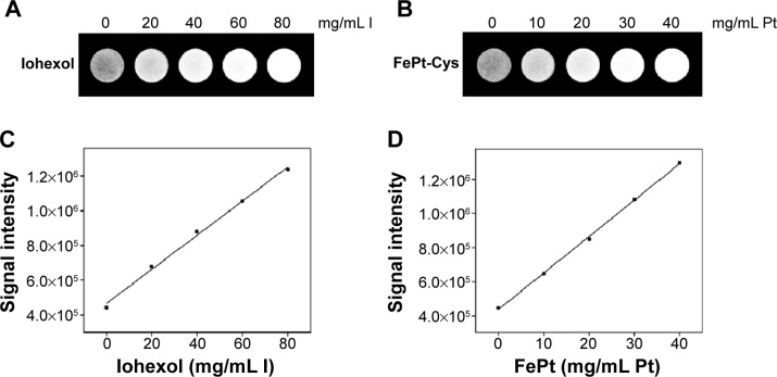
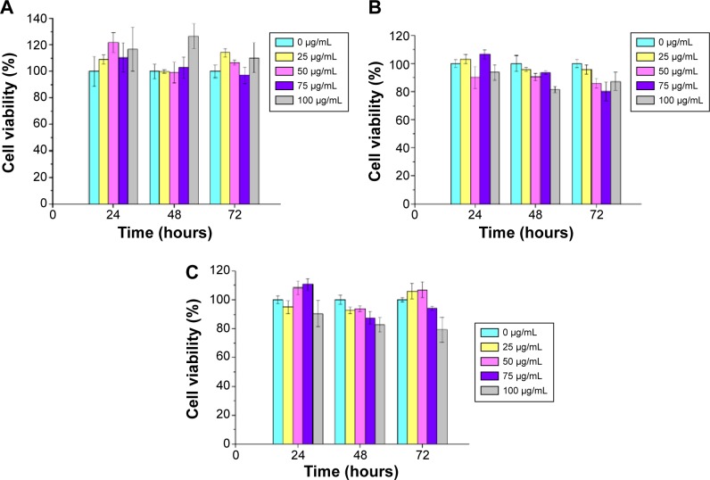
Nanoparticles (NPs) are advantageous for the delivery of diagnosis agents to brain tumors. In this study, we attempted to develop an L-cysteine coated FePt (FePt-Cys) NP as MRI/CT imaging contrast agent for the diagnosis of malignant gliomas. FePt-Cys NPs were synthesized through a co-reduction route, which was characterized by transmission electron microscopy, high-resolution transmission electron microscopy, powder X-ray diffraction, Fourier transform infrared spectroscopy, and dynamic light scattering. The MRI and CT imaging ability of FePt-Cys NPs was evaluated using different gliomas cells (C6, SGH44, U251) as the model. Furthermore, the biocompatibility of the as-synthesized FePt-Cys NPs was evaluated using three different cell lines (ECV304, L929, and HEK293) as the model. The results showed that FePt-Cys NPs displayed excellent biocompatibility and good MRI/CT imaging ability, thereby indicating promising potential as a dual MRI/CT contrast agent for the diagnosis of brain malignant gliomas.

Keywords: CT; MRI; glioma.


Figures

Figure 1
Figure 1
TEM (A) and HRTEM (B) images of as-synthesized FePt-Cys NPs. Note: The arrows in the inset of B indicate the distance between the two adjacent planes. Abbreviations: d, interplanar distance; FePt-Cys, l-cysteine coated FePt; HRTEM, high-resolution TEM; NPs, nanoparticles; TEM, transmission electron microscopy.
Figure 2
Figure 2
XRD pattern of FePt-Cys NPs. Abbreviations: FePt-Cys, l-cysteine coated FePt; NPs, nanoparticles; XRD, powder X-ray diffraction.
Figure 3
Figure 3
FT-IR spectra of pure Cys molecules (A) and FePt-Cys NPs (B). Abbreviations: Cys, l-cysteine; FePt-Cys, l-cysteine coated FePt; FT-IR, Fourier transform infrared spectroscopy; NPs, nanoparticles.
Figure 4
Figure 4
Size distributions of FePt-Cys dispersed in different media for different times. Notes: (A) Size distribution of FePt-Cys NPs dispersed in H2O. (B) Size changes of FePt-Cys NPs in H2O and cell culture media (DMEM +10% FBS) in the period of 7 days. Abbreviations: DMEM, Dulbecco’s Modified Eagle’s Medium; FBS, fetal bovine serum; FePt-Cys, l-cysteine coated FePt; NPs, nanoparticles; PDI, polydispersity index.
Figure 5
Figure 5
Magnetization hysteresis loop of the as-synthesized FePt-Cys NPs. Abbreviations: FePt-Cys, l-cysteine coated FePt; NPs, nanoparticles.
Figure 6
Figure 6
MR contrast imaging of FePt-Cys NPs. Notes: (A) The T2-weighted MR images of FePt-Cys NPs at different Fe concentrations. (B) T2 relaxation rates (1/T2) plotted against the Fe concentration of FePt-Cys NPs. Abbreviations: FePt-Cys, l-cysteine coated FePt; MR, magnetic resonance; NPs, nanoparticles; r2, relaxation rate.
Figure 7
Figure 7
CT imaging evaluation of different samples. Notes: (A) The CT images of iohexol at different iodine concentrations; (B) The CT images of FePt-Cys NPs at different Pt concentrations; (C) The signal intensity of iohexol plotted against the concentration of iodine; (D) The signal intensity of FePt-Cys NPs plotted against the concentration of Pt. Abbreviations: CT, computed tomography; FePt-Cys, l-cysteine coated FePt; NPs, nanoparticles.
Figure 8
Figure 8
Imaging of different gliomas cells (C6, SGH44, and U251) treated with FePt-Cys NPs. Notes: (A) MR imaging of different gliomas cells at different Fe concentrations; (B) CT imaging of different gliomas cells at different Pt concentrations. Abbreviations: CT, computed tomography; FePt-Cys, l-cysteine coated FePt; MR, magnetic resonance; NPs, nanoparticles.
Figure 9
Figure 9
Viabilities of different cell lines treated with FePt-Cys NPs at different Fe concentrations and time intervals. Notes: (A) ECV 304 cells; (B) L929 cells; and (C) HEK293 cells. Abbreviations: FePt-Cys, l-cysteine coated FePt; NPs, nanoparticles.

References

    1. Wen PY, Kesari S. Malignant gliomas in adults. N Engl J Med. 2008;359(5):492–509. - PubMed
    1. Orringer DA, Koo YE, Chen T, Kopelman R, Sagher O, Philbert MA. Small solutions for big problems: the application of nanoparticles to brain tumor diagnosis and therapy. Clin Pharmacol Ther. 2009;85(5):531–534. - PMC - PubMed
    1. Rozhkova EA. Nanoscale materials for tackling brain cancer: recent progress and outlook. Adv Mater. 2011;23(24):H136–H150. - PubMed
    1. Li M, Deng H, Peng H, Wang Q. Functional nanoparticles in targeting gliomas diagnosis and therapies. J Nanosci Nanotechnol. 2014;14(1):415–432. - PubMed
    1. Cheng Y, Morshed RA, Auffinger B, Tobias AL, Lesniak MS. Multifunctional nanoparticles for brain tumor imaging and therapy. Adv Drug Deliv Rev. 2014;66:42–57. - PMC - PubMed

Publication types