. 2015 Dec 15;408(2):292-304.
doi: 10.1016/j.ydbio.2015.03.013. Epub 2015 Apr 4.

ATP4a is required for development and function of the Xenopus mucociliary epidermis - a potential model to study proton pump inhibitor-associated pneumonia

Affiliations

ATP4a is required for development and function of the Xenopus mucociliary epidermis - a potential model to study proton pump inhibitor-associated pneumonia

Peter Walentek et al. Dev Biol. .

Abstract

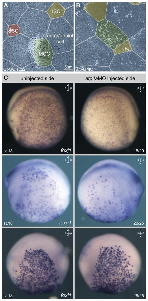
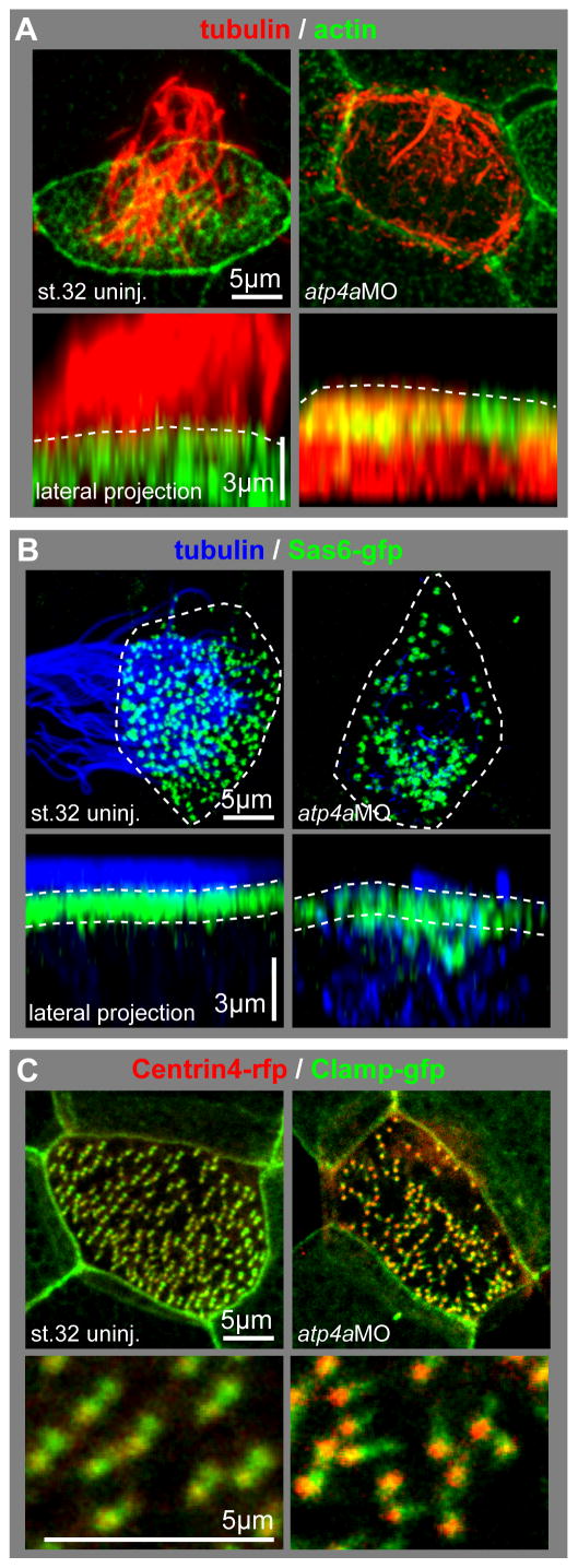
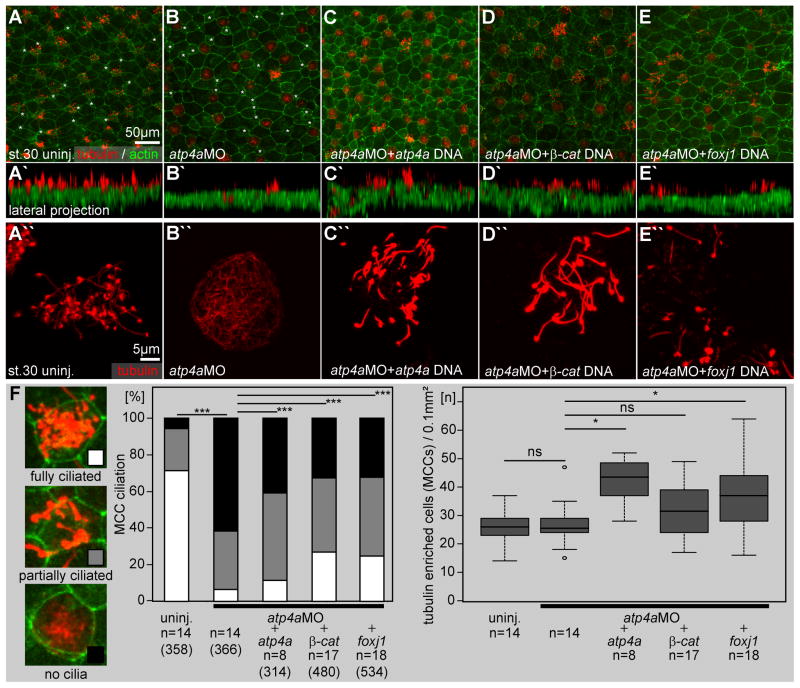
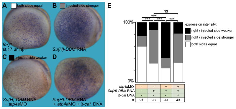
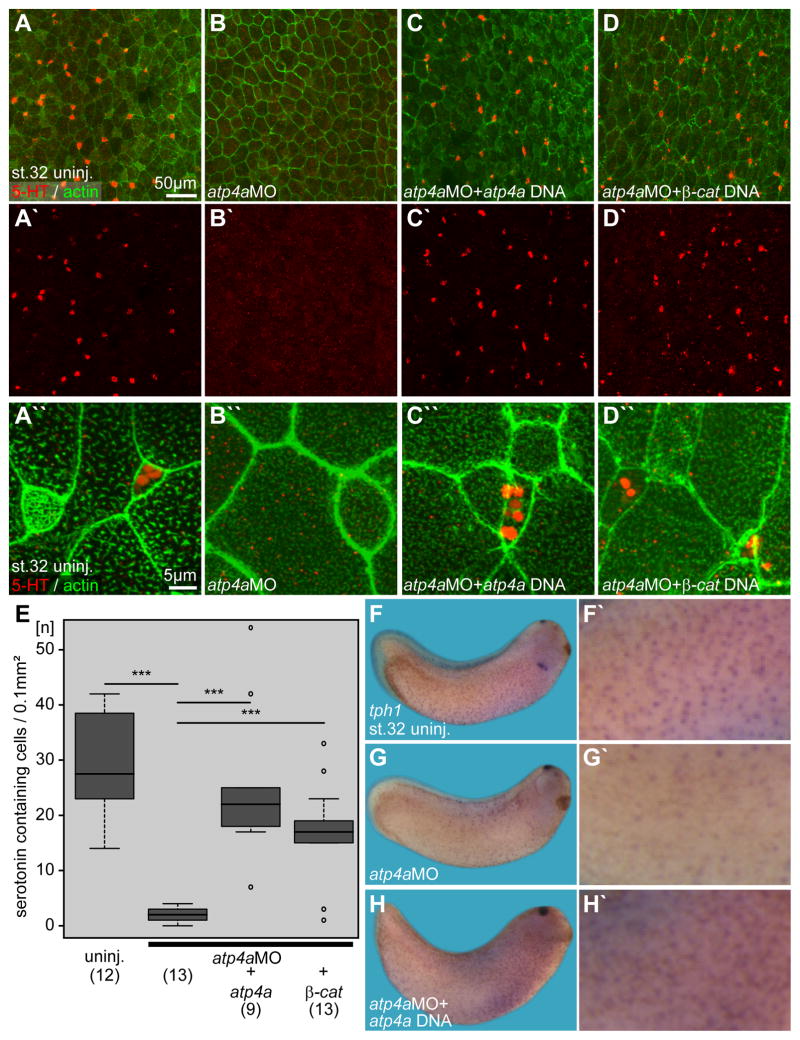
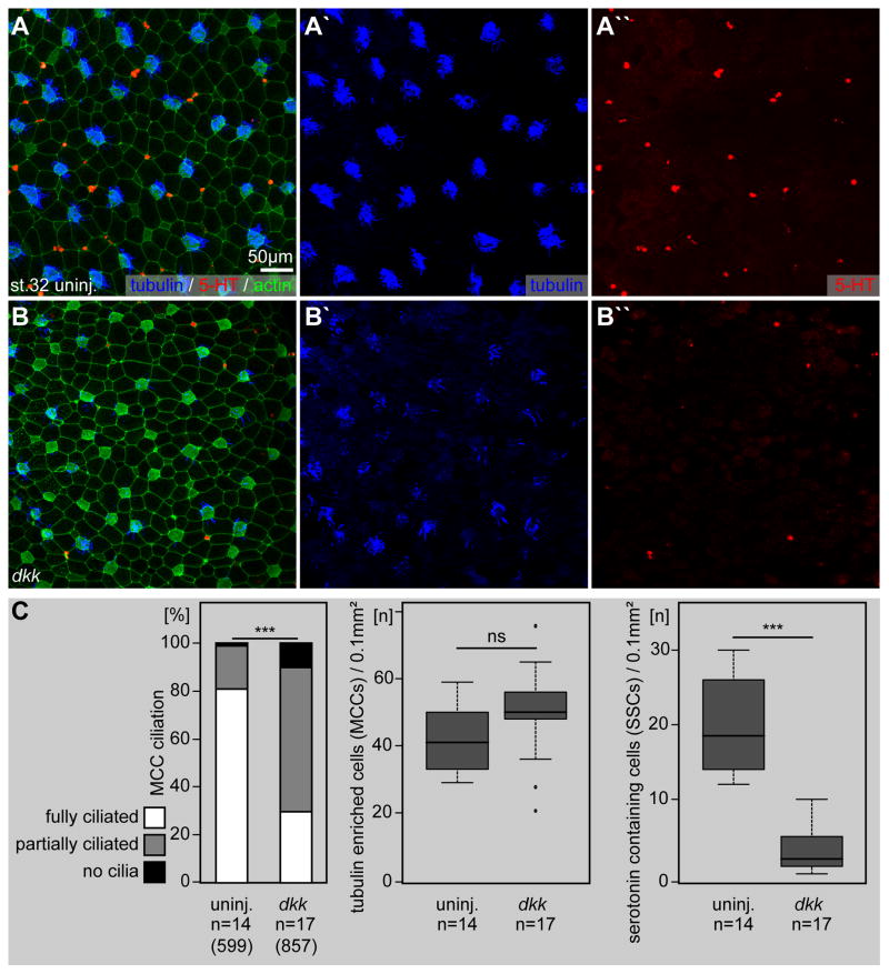
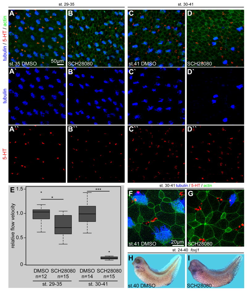
Proton pump inhibitors (PPIs), which target gastric H(+)/K(+)ATPase (ATP4), are among the most commonly prescribed drugs. PPIs are used to treat ulcers and as a preventative measure against gastroesophageal reflux disease in hospitalized patients. PPI treatment correlates with an increased risk for airway infections, i.e. community- and hospital-acquired pneumonia. The cause for this correlation, however, remains elusive. The Xenopus embryonic epidermis is increasingly being used as a model to study airway-like mucociliary epithelia. Here we use this model to address how ATP4 inhibition may affect epithelial function in human airways. We demonstrate that atp4a knockdown interfered with the generation of cilia-driven extracellular fluid flow. ATP4a and canonical Wnt signaling were required in the epidermis for expression of foxj1, a transcriptional regulator of motile ciliogenesis. The ATP4/Wnt module activated foxj1 downstream of ciliated cell fate specification. In multiciliated cells (MCCs) of the epidermis, ATP4a was also necessary for normal myb expression, apical actin formation, basal body docking and alignment of basal bodies. Furthermore, ATP4-dependent Wnt/β-catenin signaling in the epidermis was a prerequisite for foxa1-mediated specification of small secretory cells (SSCs). SSCs release serotonin and other substances into the medium, and thereby regulate ciliary beating in MCCs and protect the epithelium against infection. Pharmacological inhibition of ATP4 in the mature mucociliary epithelium also caused a loss of MCCs and led to impaired mucociliary clearance. These data strongly suggest that PPI-associated pneumonia in human patients might, at least in part, be linked to dysfunction of mucociliary epithelia of the airways.

Keywords: ATP4; Cilia; Proton pump inhibitor; Small secretory cells; Wnt; Xenopus laevis.


Figures

Fig. 1
Fig. 1. ATP4 is required for normal development of the embryonic epidermis
(A–B) Morphological analysis of the embryonic epidermis at stage 32 in control morpholino oligonucleotide (CoMO) (A) and atp4aMO (B) injected specimens. Knockdown of atp4a lead to defects in ciliation of multiciliated cells (MCCs, green) and reduced numbers of small secretory cells (SSCs, red), but without obvious effects on ion secreting cells (ISCs, yellow) or outer/goblet cells (blue). (C) Knockdown of atp4a attenuated foxj1 and foxa1 expression, but not foxi1 expression in the skin epidermis. Embryos were unilaterally injected with atp4aMO at the four-cell stage and assayed for foxj1/foxa1/foxi1 expression by WMISH at stage 15. Correct targeting was confirmed by co-injection of lineage tracer. Depicted embryos are derived from the same injected batch. Numbers in the right lower corner indicate frequency of phenotype. a, anterior; d, dorsal; p, posterior; st., stage; v, ventral.
Fig. 2
Fig. 2. Loss of ATP4 causes defects in MCCs reminiscent of foxj1 and Wnt/PCP phenotypes
(A–C) Analysis of the cellular phenotype in MCCs using high magnification confocal single cell imaging. (A) Cells were stained with an antibody against acetylated-α-tubulin (tubulin, red) and phalloidin-Alexa488 for actin staining (actin, green). Control (uninj.) MCCs were characterized by the presence of a dense ciliary tuft projecting from the apical surface (dashed lines in lateral projections) and the presence of an apical actin meshwork. In contrast, MCCs in atp4a morphants showed reduced ciliation, intracellular accumulation of tubulin and defects in the apical actin meshwork formation. (B) The localization of basal bodies to the apical membrane was analyzed using overexpression of sas6-gfp (green) in combination with tubulin (blue) staining, the outlines of the cells are depicted by the dashed lines in the apical views. Control MCCs were fully ciliated and basal bodies aligned close to the apical membrane (dashed lines in lateral projections), while atp4a morphant MCCs had severe defects in ciliation, which correlated with aberrant basal body distribution within the cell. Basal bodies in atp4a morphants were mislocalized to the deep cytoplasm and not uniformly distributed along the apical membrane. (C) Basal body orientation was analyzed using centrin4-rfp (red) and clamp-gfp (green) overexpression. In control MCCs, basal bodies were mostly uniformly aligned along the anterior-posterior axis, while this alignment was randomized in atp4a morphant embryos. Lower panels show a magnified field of basal bodies.
Fig. 3
Fig. 3. ATP4-dependent Wnt signaling is required for foxj1 expression and ciliation in the Xenopus embryonic skin
(A–E) Immunofluorescent analysis of skin ciliation at stage 30. Embryos were stained for tubulin (acetylated-α-tubulin, red) and actin (phalloidin-Alexa488, green) and analyzed by confocal microscopy. (B) In atp4a morphants fully ciliated, partially ciliated and non-ciliated MCCs were found. Smaller intercalating cells with ISC morphology, which are also negative for acetylated-α-tubulin staining are marked with asterisks in A and B. Note that atp4a (C), β-catenin (β-cat.; D) and foxj1 (E) DNAs partially rescued ciliation in atp4a morphants. (A′–E′) Lateral projections of confocal Z-scans are shown in (A–E). (A″–E″) Higher magnification of representative MCCs. (F) Quantification of results. n, number of embryos; (n), number of cells.
Fig. 4
Fig. 4. ATP4a-dependent Wnt signaling acts downstream of Notch in skin foxj1 induction
(A–D) Foxj1 was stained in uninjected control (uninj.) embryos (A), as well as manipulated embryos (injected side shown in B–D). (B) Inhibition of Notch by injection of Su(H)-DBM mRNA increased foxj1 expression, which remained dependent on ATP4a (C) and Wnt/β-catenin (β-cat.; D). (E) Quantification of results. Staining intensity on the injected (right) side was compared to the uninjected (left) control side and quantified as right/injected side stronger, weaker or equal to the control side (G). a, anterior; d, dorsal; n, number of embryos; ns, not significant; p, posterior; st., stage; v, ventral.
Fig. 5
Fig. 5. Serotonin secretion and tph1 expression in the embryonic epidermis are regulated by ATP4a-mediated Wnt/β-catenin signaling
(A–D) Immunofluorescent analysis and quantification (E) of serotonin (5-HT; red) deposition in small secretory cells (SSCs) of the skin at stage 32. Actin (phalloidin-Alexa488) staining in green. (A) Uninjected (uninj.) control. (B) Loss of serotonin staining in atp4a morphants. (C, D) Rescue of serotonin staining upon co-injection of atp4a (C) or β-catenin (β-cat.; D) DNA constructs. (A′–D′) Serotonin channel. (A″–D″) Higher magnification of (A–D). (E) Quantification of results. (F–H) WMISH for tph1. (F, F′) Uninjected control. (G, G′) Loss of tph1 expression in atp4a morphants was rescued by co-injection of atp4a DNA (H). n, number of embryos; st., stage.
Fig. 6
Fig. 6. Endogenous Wnt/β-catenin signaling is required for ciliation and serotonin signaling in the ciliated epidermis
Immunofluorescent analysis (A, B) and quantification (C) of MCC ciliogenesis (acetylated-α-tubulin, blue) and serotonin (5-HT; red) deposition in small secretory cells (SSCs) of the skin at stage 32. Actin (phalloidin-Alexa488) staining in green. (A) Uninjected (uninj.) controls are characterized by dense ciliation of MCCs (A′) and the presence of large numbers of serotonin positive SSCs (A″). (B) Impaired ciliogenesis and loss of serotonin staining in dkk injected specimens. dkk injected embryos showed a decrease in ciliation in MCCs (B′) and reduced numbers serotonin stained cells (B″). n, number of embryos; (n), number of cells; st., stage.
Fig. 7
Fig. 7. Late pharmacological inhibition of ATP4 causes loss of MCCs and decreased mucociliary clearance
Immunofluorescent analysis (A–D) of MCC ciliogenesis (acetylated-α-tubulin, blue) and serotonin (5-HT; red) deposition in small secretory cells (SSCs) of the skin at stages 35 (AB) and 41 (C–D). Actin (phalloidin-Alexa488) staining in green. Embryos were treated with DMSO as control or SCH28080 during the indicated stages. Stage 35 (A) and stage 41 (C) DMSO treated controls displayed fully ciliated MCCs (A′, C′) and the presence of large numbers of serotonin positive SSCs (A″, C″). At stage 35 no apparent defects in MCC ciliation (B′) or SSC serotonin deposition (B″) were observed in SCH28080 treated embryos (cf. Quantification in Fig. S3K), although 50% of specimens contained MCCs with a relatively small apical surface area and 60% of specimens were abnormal in their epithelial morphology, i.e. enlarged cells were present (cf. Quantification in Fig. S3K). In contrast, SCH28080 treatment until stage 41 lead to a massive loss of MCCs (D′), but no effect on SSCs (D″). (E) Quantification of cilia-driven fluid flow velocities revealed a significantly reduced extracellular fluid flow at the epidermis at stages 35 and 41, as compared to DMSO treated controls of the same stage. (F–G) High-magnification confocal imaging on the same set of specimens depicted in (C and D) confirmed the lack of MCCs, but intact SSCs in SCH28080 treated embryos and further indicated apical expansion in ISC-like cells and SSCs as well as enriched actin staining at some cell junctions (G). MCCs and epithelial morphology appeared normal in DMSO treated controls (F). (H–I) Embryos were treated with DMSO or SCH28080 from stage 24–40 and foxj1 expression was analyzed by in situ hybridization. In comparison to controls (H), foxj1 staining was increased in SCH28080 treated tadpoles (I). n, number of embryos; st., stage.

Similar articles

Cited by

References

    1. Altman KW, Waltonen JD, Tarjan G, Radosevich Ja, Haines GK. Human lung mucous glands manifest evidence of the H+/K+-ATPase proton pump. Ann Otol Rhinol Laryngol. 2007;116:229–34. - PubMed
    1. Avasthi P, Marshall WF. Stages of ciliogenesis and regulation of ciliary length. Differentiation. 2012;83:S30–42. doi: 10.1016/j.diff.2011.11.015. - DOI - PMC - PubMed
    1. Bayer H, Müller T, Myrtek D, Sorichter S, Ziegenhagen M, Norgauer J, Zissel G, Idzko M. Serotoninergic receptors on human airway epithelial cells. Am J Respir Cell Mol Biol. 2007;36:85–93. doi: 10.1165/rcmb.2006-0151OC. - DOI - PubMed
    1. Bergmann C. Educational paper: ciliopathies. Eur J Pediatr. 2012;171:1285–300. doi: 10.1007/s00431-011-1553-z. - DOI - PMC - PubMed
    1. Beyer T, Danilchik M, Thumberger T, Vick P, Tisler M, Schneider I, Bogusch S, Andre P, Ulmer B, Walentek P, Niesler B, Blum M, Schweickert A. Serotonin Signaling Is Required for Wnt-Dependent GRP Specification and Leftward Flow in Xenopus. Curr Biol. 2012;22:1–7. doi: 10.1016/j.cub.2011.11.027. - DOI - PubMed

Publication types

MeSH terms