. 2015 Apr 7;10(4):e0122992.
doi: 10.1371/journal.pone.0122992. eCollection 2015.

High expression of KCa3.1 in patients with clear cell renal carcinoma predicts high metastatic risk and poor survival

Affiliations

High expression of KCa3.1 in patients with clear cell renal carcinoma predicts high metastatic risk and poor survival

Maj Rabjerg et al. PLoS One. .

Abstract

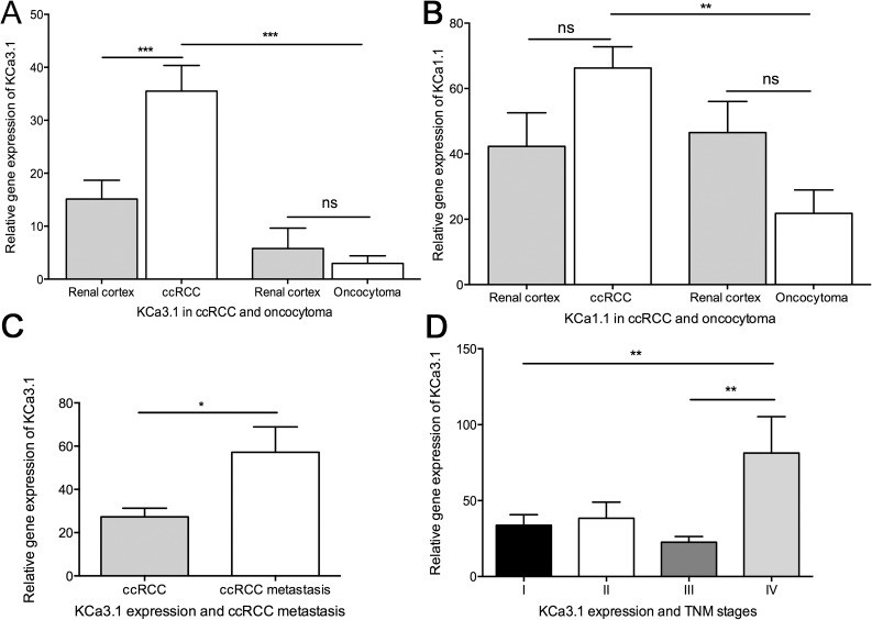
Background: Ca2+-activated K+ channels have been implicated in cancer cell growth, metastasis, and tumor angiogenesis. Here we hypothesized that high mRNA and protein expression of the intermediate-conductance Ca2+-activated K+ channel, KCa3.1, is a molecular marker of clear cell Renal Cell Carcinoma (ccRCC) and metastatic potential and survival.

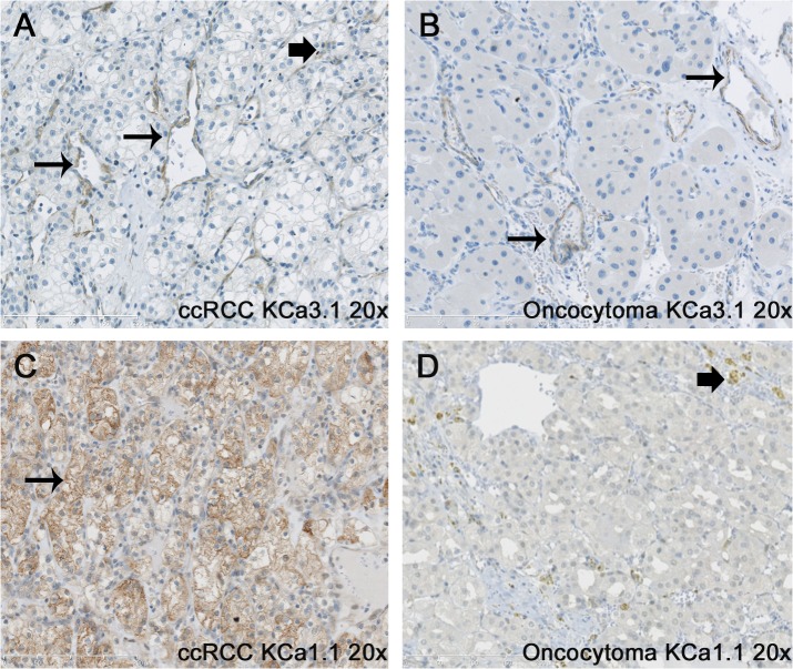
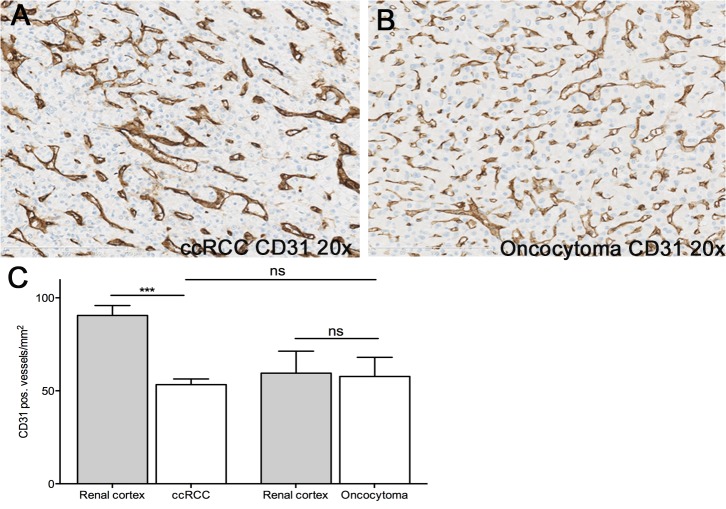
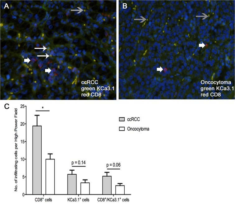
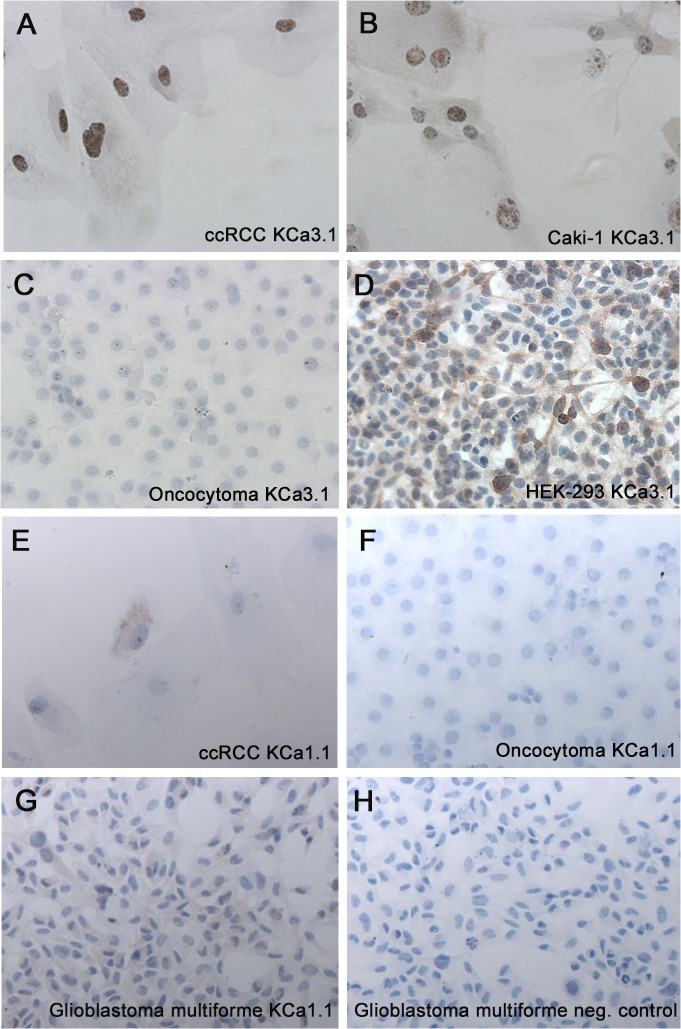
Methodology/principal findings: We analyzed channel expression by qRT-PCR, immunohistochemistry, and patch-clamp in ccRCC and benign oncocytoma specimens, in primary ccRCC and oncocytoma cell lines, as well as in two ccRCC cell lines (Caki-1 and Caki-2). CcRCC specimens contained 12-fold higher mRNA levels of KCa3.1 than oncocytoma specimens. The large-conductance channel, KCa1.1, was 3-fold more highly expressed in ccRCC than in oncocytoma. KCa3.1 mRNA expression in ccRCC was 2-fold higher than in the healthy cortex of the same kidney. Disease specific survival trended towards reduction in the subgroup of high-KCa3.1-expressing tumors (p<0.08 vs. low-KCa3.1-expressing tumors). Progression-free survival (time to metastasis/recurrence) was reduced significantly in the subgroup of high-KCa3.1-expressing tumors (p<0.02, vs. low-KCa3.1-expressing tumors). Immunohistochemistry revealed high protein expression of KCa3.1 in tumor vessels of ccRCC and oncocytoma and in a subset of ccRCC cells. Oncocytoma cells were devoid of KCa3.1 protein. In a primary ccRCC cell line and Caki-1/2-ccRCC cells, we found KCa3.1-protein as well as TRAM-34-sensitive KCa3.1-currents in a subset of cells. Furthermore, Caki-1/2-ccRCC cells displayed functional Paxilline-sensitive KCa1.1 currents. Neither KCa3.1 nor KCa1.1 were found in a primary oncocytoma cell line. Yet KCa-blockers, like TRAM-34 (KCa3.1) and Paxilline (KCa1.1), had no appreciable effects on Caki-1 proliferation in-vitro.

Conclusions/significance: Our study demonstrated expression of KCa3.1 in ccRCC but not in benign oncocytoma. Moreover, high KCa3.1-mRNA expression levels were indicative of low disease specific survival of ccRCC patients, short progression-free survival, and a high metastatic potential. Therefore, KCa3.1 is of prognostic value in ccRCC.


Conflict of interest statement

Competing Interests: The authors have declared that no competing interests exists.

Figures

Fig 1
Fig 1. Quantitative RT-PCR analysis of KCa3.1 and KCa1.1 mRNA in oncocytoma and ccRCC together with paired normal renal cortex.
Mean + SEM is shown. KCa3.1 gene expression is shown relative to reference gene expression of multiple reference genes and KCa1.1 gene expression relative to reference gene TBP. A) Comparison of KCa3.1 gene expression in tumor tissue from oncocytoma and ccRCC together with paired unaffected renal cortex, B) Comparison of KCa1.1 gene expression in tumor tissue from oncocytoma and ccRCC together with paired unaffected renal cortex C) KCa3.1 gene expression in ccRCC tumors with and without metastasis, D) Comparison of KCa3.1 gene expression in different TNM stages of ccRCC. *p < 0.05, **p< 0.01 ***p < 0.001. ns = non-significant.
Fig 2
Fig 2. KCa3.1-mRNA expression levels are a significant prognostic indicator of Progression Free Survival in ccRCC.
A-C: Kaplan Meier survival analysis indicates that patients with high KCa3.1-mRNA expression have a significantly shorter Progression Free Survival (p = 0.02). Moreover, we found a trend towards a significantly shorter Disease Specific Survival for patients with a high KCa3.1 mRNA expression (p = 0.08).
Fig 3
Fig 3. Immunohistochemical staining for KCa3.1 in ccRCC and oncocytoma.
Immunohistochemical staining for KCa3.1 in ccRCC (A) and oncocytoma (B) shows strong staining of tumor vessels (long arrows) in both tumor subtypes. In ccRCC, a few tumor cells or possibly stroma cells show some KCa3.1 expression (“block” arrow). Immunohistochemical staining for KCa1.1 in ccRCC (C) and oncocytoma (D) shows staining of the cell membrane of the tumor clear cells (long arrow), whereas no staining of the tumor cells was observed in the oncocytoma. “Block” arrow indicates staining of immune cells. Original magnification, x200.
Fig 4
Fig 4. Immunohistochemical staining for CD31 in ccRCC and oncocytoma.
Immunohistochemical staining for CD31 in ccRCC (A) and oncocytoma (B) showed no difference in microvessel density between the two tumor subtypes (C). Note that the unaffected cortex from ccRCC patients contained more CD31-positive blood vessels while the number of vessels was alike in oncocytoma and paired cortex. Data are given as mean ± SEM. Number of patients: n = 7; *** p<0.001 vs. ccRCC, One-way ANOVA followed by Tukey post hoc test.
Fig 5
Fig 5. Immunofluorescent staining for CD8 and KCa3.1 in ccRCC and oncocytoma.
Immunofluorescence of CD8 (red) and KCa3.1 (green) in ccRCC (A) and oncocytoma (B). Large arrows indicate T cells positive for CD8 and KCa3.1. Small arrows indicate T cells positive for CD8 but KCa3.1-negative. Grey arrows indicate erythrocytes within tumor vessels that exhibited staining for KCa3.1 and CD8 that could be, however, auto-fluorescence. (C) Quantification of CD8-positive T cells, KCa3.1-positive CD8 T cells, and other cells in ccRCC and oncocytoma tumor tissues. Data are given as mean ± SEM. Number of tumors: n = 7, each; * p<0.05 ccRCC vs. oncocytoma, One-way ANOVA followed by Tukey post hoc test.
Fig 6
Fig 6. Immunocytochemical staining for KCa3.1 in a ccRCC cell line.
Immunohistochemical staining for KCa3.1 in a primary ccRCC cell line showed relatively weak and heterogeneous membrane staining and an intense staining of presumably the endoplasmic reticulum around nuclei (A). Similar pattern of KCa3.1-staining was seen in Caki-1 cells (B), while primary oncocytoma cells lacked KCa3.1-stain (C). KCa3.1-transfected HEK cells served as a positive control (D). Immunohistochemical staining for KCa1.1 in primary ccRCC showed a weak staining of the membrane (E), whereas no staining was observed in the primary oncocytoma (F). A glioblastoma cell line (U251 MG) served as positive control for KCa1.1 (G-H). Original magnification, 200x.
Fig 7
Fig 7. Positive KCa3.1 currents detected in ccRCC by patchclamping.
(A) Electrophysiological characterization of KCa3.1 channels in ccRCC. In primary ccRCC, KCa3.1 whole-cell currents exhibited typical KCa3.1 features such as inward rectification at positive voltage and complete inhibition by the selective KCa3.1 blocker, TRAM-34. KCa3.1 currents were not detected in any of the oncocytoma cells. Caki cells displayed consistently TRAM-34-sensitive KCa3.1 currents. The graph shows summary data of KCa3.1 current densities and blockade of KCa3.1 by TRAM-34. (B) Electrophysiological characterization of KCa1.1 channel in ccRCC. In primary ccRCC, we saw a KCa1.1-typical voltage-dependent I/U relationship with large current amplitudes at positive membrane potentials. KCa1.1 currents were large in ccRCC, sensitive to Paxilline, and virtually undetectable in oncocytoma. Data are mean ± SEM. Number in graph indicate the number of repetitions; *p< 0.05.

Similar articles

Cited by

References

    1. Ljungberg B, Campbell SC, Choi HY, Jacqmin D, Lee JE, Weikert S et al. (2011) The epidemiology of renal cell carcinoma. Eur Urol 60: 615–621. 10.1016/j.eururo.2011.06.049 - DOI - PubMed
    1. Ishii TM, Silvia C, Hirschberg B, Bond CT, Adelman JP, Maylie J et al. (1997) A human intermediate conductance calcium-activated potassium channel. Proc Natl Acad Sci U S A 94: 11651–11656. - PMC - PubMed
    1. Wei AD, Gutman GA, Aldrich R, Chandy KG, Grissmer S, Wulff H et al. (2005) International Union of Pharmacology. LII. Nomenclature and molecular relationships of calcium-activated potassium channels. Pharmacol Rev 57: 463–472. - PubMed
    1. Wulff H, Kolski-Andreaco A, Sankaranarayanan A, Sabatier JM, Shakkottai V (2007) Modulators of small- and intermediate-conductance calcium-activated potassium channels and their therapeutic indications. Curr Med Chem 14: 1437–1457. - PubMed
    1. Thompson-Vest N, Shimizu Y, Hunne B, Furness JB (2006) The distribution of intermediate-conductance, calcium-activated, potassium (IK) channels in epithelial cells. J Anat 208: 219–229. - PMC - PubMed

Publication types

Substances