. 2015 Feb 18:15:56.
doi: 10.1186/s12870-014-0362-8.

Antibody-based screening of cell wall matrix glycans in ferns reveals taxon, tissue and cell-type specific distribution patterns

Antibody-based screening of cell wall matrix glycans in ferns reveals taxon, tissue and cell-type specific distribution patterns

Olivier Leroux et al. BMC Plant Biol. .

Abstract

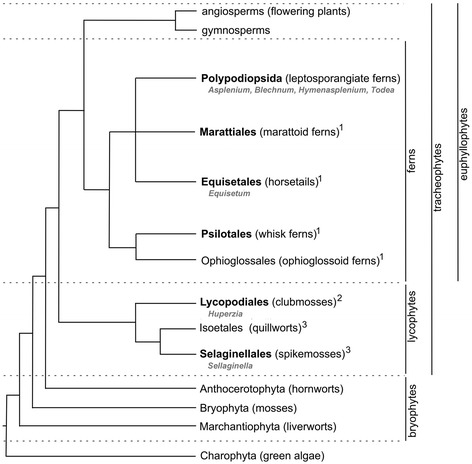
Background: While it is kno3wn that complex tissues with specialized functions emerged during land plant evolution, it is not clear how cell wall polymers and their structural variants are associated with specific tissues or cell types. Moreover, due to the economic importance of many flowering plants, ferns have been largely neglected in cell wall comparative studies.

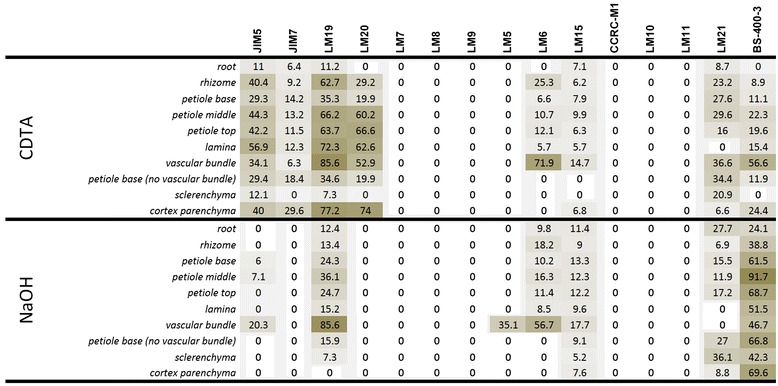
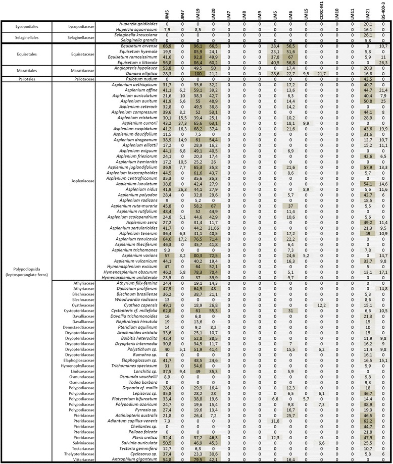
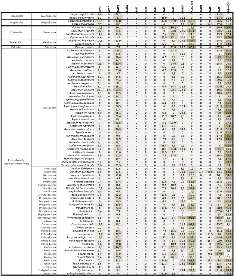
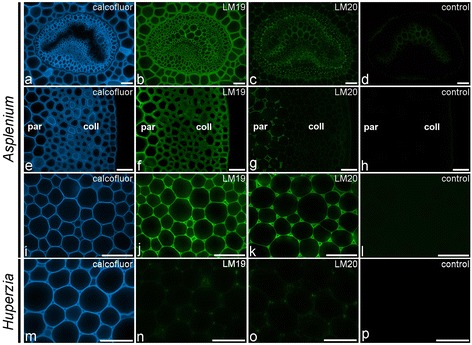
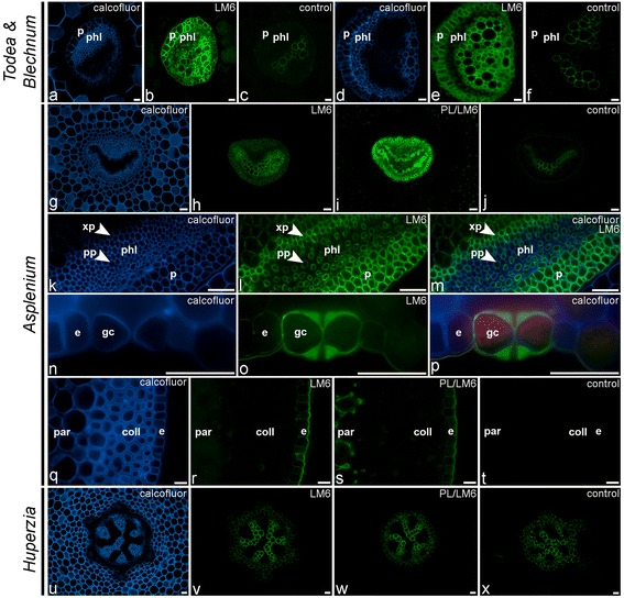
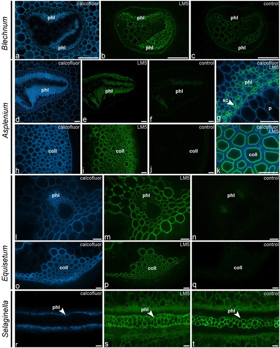
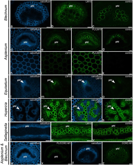
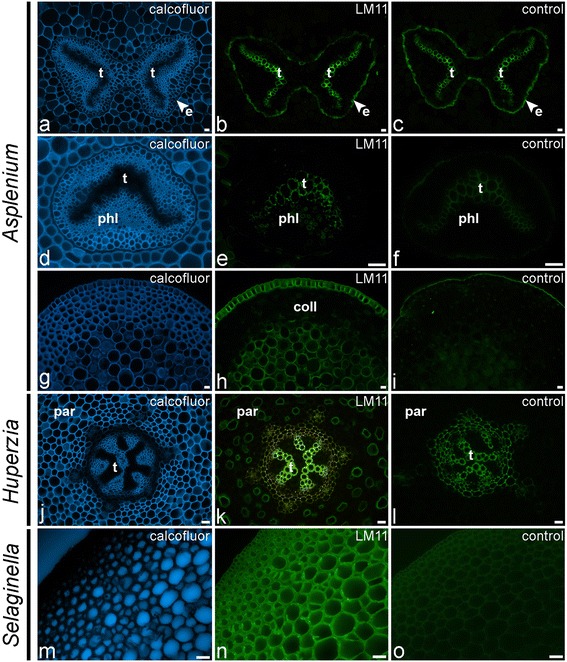
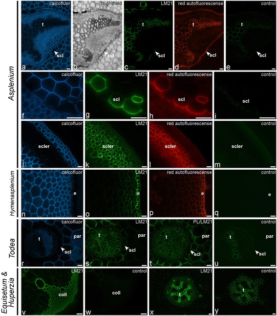
Results: To explore fern cell wall diversity sets of monoclonal antibodies directed to matrix glycans of angiosperm cell walls have been used in glycan microarray and in situ analyses with 76 fern species and four species of lycophytes. All major matrix glycans were present as indicated by epitope detection with some variations in abundance. Pectic HG epitopes were of low abundance in lycophytes and the CCRC-M1 fucosylated xyloglucan epitope was largely absent from the Aspleniaceae. The LM15 XXXG epitope was detected widely across the ferns and specifically associated with phloem cell walls and similarly the LM11 xylan epitope was associated with xylem cell walls. The LM5 galactan and LM6 arabinan epitopes, linked to pectic supramolecules in angiosperms, were associated with vascular structures with only limited detection in ground tissues. Mannan epitopes were found to be associated with the development of mechanical tissues. We provided the first evidence for the presence of MLG in leptosporangiate ferns.

Conclusions: The data sets indicate that cell wall diversity in land plants is multifaceted and that matrix glycan epitopes display complex spatio-temporal and phylogenetic distribution patterns that are likely to relate to the evolution of land plant body plans.


Figures

Figure 1
Figure 1
Schematic tree showing the relationships among the major groups of land plants. 1: eusporangiate ferns s.l.; 2: homosporous lycophytes; 3: heterosporous lycophytes. Representatives of the plant groups indicated in bold were sampled for this study (see Supplementary Figure 1). Genera represented in the immunofluorescence figures are indicated (grey). Adapted from [74,75].
Figure 2
Figure 2
Glycan microarray heatmap of CDTA and NaOH extracts of total organ or isolated tissue(s) of the leptosporangiate fern Asplenium elliottii . The probes are listed at the top of the heatmap. References for probe specificity are listed in Table 1. Abbreviations: mAb: monoclonal antibody; HG: pectic homogalacturonan; AGP: arabinogalactan protein; XG: xyloglucan; Me: methyl-esterified.
Figure 3
Figure 3
Glycan microarray heatmap of CDTA extracts of fern or lycophyte petioles/stems. References for probe specificity are listed in Table 1.
Figure 4
Figure 4
Glycan microarray heatmap of NaOH extracts of fern or lycophyte petioles/stems. References for probe specificity are listed in Table 1.
Figure 5
Figure 5
Indirect immunofluorescence detection of homogalacturonan epitopes with low (LM19) and high (LM20) levels of esterification in fern petioles and lycophyte stems. Calcofluor White fluorescence (a, e, i, m) shows the full extent of cell walls. (a-d) LM19 is detected in primary cell walls of the vascular bundle of Asplenium rutifolium, while LM20 is restricted to the middle lamellae and intercellular space corners. (e–h) The prevalence of the LM19 epitope over the LM20 epitope is apparent in parenchymatous and collenchymatous tissue of Asplenium rutifolium. (i–l) LM19 is detected in primary cell walls of Asplenium daucifolium, while LM20 is restricted to the middle lamellae and intercellular space corners. (m–p) LM19 and LM20 weakly bind to primary cell walls in the lycophyte Huperzia squarrosum. Abbreviations: par, parenchyma; coll, collenchymatous tissue. No primary antibody controls are provided (d, h, l, p). Scale bars: 40 μm.
Figure 6
Figure 6
Indirect immunofluorescence detection of the arabinan (LM6) epitope in transverse sections of fern petioles and lycophyte stems. Calcofluor White fluorescence (a, d, g, k, m, n, p, q, u) shows the full extent of cell walls. (a–c) Detection of the LM6-epitope in parenchymatous cell types of vascular bundles of Todea sp. (a–c) and Blechnum brasiliense (d–f). (g–m) Similar distribution pattern of the LM6-epitope is found in the vascular bundle (g–j) of Asplenium theciferum. Higher magnification (k–m) showing binding of LM6 to the cell walls of phloem parenchyma (pp), xylem parenchyma (xp) and pericycle (p). (n–p) LM6 binding to epidermal (e) cell walls, including the guard cell walls (gc) of stomata. (q–t) Detection of LM6 epitope in cortical parenchyma after pectate lyase (PL) treatment (s). (u–x) LM6-epitope is not detected in the lycophyte Huperzia squarrosum, even after pectate lyase treatment (w). Abbreviations: p, pericycle; phl, phloem; xp, xylem parenchyma; pp, phloem parenchyma; par, parenchyma; coll, collenchymatous tissue; e, epidermis. No primary antibody controls are provided (c, f, j, t, x). Scale bars: 40 μm.
Figure 7
Figure 7
Indirect immunofluorescence detection of the galactan (LM5) epitope in transverse sections of fern petioles and lycophyte stems. Calcofluor White fluorescence (a, d, g, h, k, l, o, r) shows the full extent of cell walls. (a–c) Abundance of the LM5 epitope in cell walls of phloem sieve cells in the vascular bundle of Blechnum brasiliense. (d–g) Similar distribution pattern of LM5 in Asplenium compressum. A high magnification (g) of a vascular bundle shows that LM5-binding is restricted to cell walls of phloem sieve cells (sc). (h–k) Binding of LM5 to the innermost cell wall layers of collenchymatous tissue in Asplenium rutifolium. (l–q) LM5 binding to most tissues in Equisetum arvense, including the cell walls of the vascular bundle and surrounding parenchyma (l–n) as well as to the inner cell wall layer of the collenchymatous strengthening tissue (o–q). (r–t) LM5 binding to phloem in the lycophyte Selaginella grandis. Abbreviations: phl, phloem; sc, sieve cell; coll, collenchymatous tissue. No primary antibody controls are provided (c, f, j, n, q, t). Scale bars: 40 μm.
Figure 8
Figure 8
Indirect immunofluorescence detection of xyloglucan (LM15, CCRC-M1) epitopes in transverse sections of fern petioles and lycophyte stems. Calcofluor White fluorescence (a, d, h, l, n, p, r, t, w, y) shows the full extent of cell walls. (a–c) Binding of LM15 to phloem cell walls of Blechnum brasiliense. (d–k) Pectate lyase treatment (PL) unmasks LM15-epitopes in phloem cell walls of Asplenium rutifolium (d–g) and in primary cell walls of cortical parenchyma of Asplenium elliottii (h–k). (l–o) LM15 binds to phloem tissue of Equisetum ramosissimum. (p–v) Similar distribution patterns are found in the lycophytes Huperzia squarrosum (p–s) and Selaginella grandis (t–v). (w–z) While the CCRC-M1 epitope is not detected in Asplenium rutifolium (w, x), even after pectate lyase pre-treatment (PL), it is localized in the phloem of Blechnum brasiliense (y, z). No primary antibody controls are provided (c, g, k, o, s, v). Abbreviations: phl: phloem. Scale bars: 40 μm.
Figure 9
Figure 9
Indirect immunofluorescence detection of the xylan (LM11) epitope in transverse sections of fern petioles and lycophyte stems. Calcofluor White fluorescence (a, d, g, j, m) shows the full extent of cell walls. (a–c) Autofluorescence of tracheids (t) obscures observation of LM11 binding in Asplenium polyodon. Note also the autofluorescence of the suberin lamellae of the endodermis (e). (d–f) LM11 binding to tracheid cell walls and phloem cell walls of Asplenium loxoscaphoides. (g–i) Abundance of the LM11 epitope in epidermal cell walls and cortical parenchyma of Asplenium rutifolium. Note that the epitope is not detected in the collenchymatous tissue (coll). (j–l) Binding of LM11 to cell walls of scattered cortical parenchyma cells in the lycophyte Huperzia squarrosum. (m–o) In the lycophyte Selaginella grandis, the LM11 epitope is detected in all cortical parenchyma cell walls. Abbreviations: t, tracheids; e, epidermis; phl, phloem; coll, collenchymatous tissue. No primary antibody controls are provided (c, f, i, l, o). Scale bars: 40 μm.
Figure 10
Figure 10
Indirect immunofluorescence detection of mannan (LM21) epitopes in transverse sections of fern petioles and lycophyte stems. Calcofluor White fluorescence (a, f, j, n, r) and bright-field (b) showing the full extent of cell walls. (a–i) Localisation of the LM21 epitope in and around the vascular bundle of Asplenium elliottii. The epitope is detected in the cell walls of tracheids (t) and sclereids (scl) that surround the vascular bundle. A high magnification shows detection of the LM21 epitope in the inner cell wall layer of sclereids (f–i). Note red autofluorescence of sclereid cell walls (d, h). (j–m) LM21 binding to the innermost cell wall layer of sclerenchyma hypodermal cells (hyp) and the sclereids of an Asplenium elliottii petiole. Note red autofluorescence of sclereid cell walls (l). (n–q) Immunodetection of LM21 in sclerified epidermal cell walls and subepidermal tissue of Hymenasplenium obscurum. Note red autofluorescence of sclerified epidermis and subepidermal tissue (p). (r–u) Unmasking of the LM21 epitope in primary cell walls of the cortical parenchyma of Todea sp. after pectate lyase treatment (PL). Note that cell walls of the vascular bundle and surrounding sclereids (scl) are labeled prior to pectate lyase treatment. (v, w) Detection of the LM21 epitope in the collenchymatous strengthening tissue of Equisetum ramossisimum. (x, y) Strong binding of LM21 to tracheids and weak binding to cortical parenchyma in the lycophyte Huperzia squarrosum. Abbreviations: scl: sclerenchyma; phl: phloem; ep: epidermis. Abbreviations: t, tracheids; scl, sclereids; scler, sclerified tissue; e, epidermis; par, parenchyma, coll, collenchymatous tissue. No primary antibody controls are provided (e, i, m, q, u, w, y). Scale bars: 40 μm.
Figure 11
Figure 11
Indirect immunofluorescence detection of the mixed-linkage glucan epitope (BioSupplies 400–3) in transverse sections of fern petioles and lycophyte stems. Calcofluor White fluorescence (a, d, g, k, o, s, v, w) and bright-field (m) showing the full extent of cell walls. (a–n) Localisation of the MLG epitope in Asplenium elliottii. The epitope is detected in collenchymatous (d–f) and sclerenchymatous (g–j) mechanical tissues as well as in epidermal cell walls. Parenchyma shows differential labelling intensities with stronger labelling of parenchyma walls surrounding mechanical tissues. Increased labelling is observed in parenchyma tissues bordering the hypodermal sclerenchyma (g–j) and the zone with sclereids surrounding the vascular bundle (k–n). Weak labelling is observed in the phloem tissue. (o–r) The anti-MLG antibody binds to parenchymatous cell walls in Blechnum brasiliense. Cell walls of the sclerenchyma sheath (ss) and subepidermal sclerenchyma (scler) are not labelled. (s–w) Detection of the MLG epitope in the collenchymatous strengthening tissue (coll) of Equisetum arvense. Higher magnification (v, w) shows that the epitope is restricted to secondary cell walls. (x, y) Labelling of a continuous ring of parenchyma tissue located between the central cavity and vascular bundles. Abbreviations: coll, collenchymatous tissue; scler, sclerenchyma; e, epidermis; scl: sclereïds; t, tracheids; ss, sclerenchyma sheath; vb: vascular bundle. No primary antibody controls are provided (f, j, n, r, u, y). Scale bars: ac, 1 mm; dw, 40 μm.

References

    1. Bateman RM, Crane PR, SiMichele WA, Kenrick PR, Rowe NP, Speck T, Stein EW. Early evolution of land plants: phylogeny, physiology, and ecology of the primary terrestrial radiation. Ann Rev Ecol System. 1998;29:263–292. doi: 10.1146/annurev.ecolsys.29.1.263. - DOI
    1. Evert RF. Esau's plant anatomy. Meristems, cells, and tissues of the plant body: their structure, function, and development. New Jersey: John Wiley and Sons, Inc; 2006.
    1. Burton RA, Gidley MJ, Fincher GB. Heterogeneity in the chemistry, structure and function of plant cell walls. Nat Chem Biol. 2010;6:724–732. doi: 10.1038/nchembio.439. - DOI - PubMed
    1. Cosgrove DJ. Growth of the plant cell wall. Nat Rev Mol Cell Biol. 2005;6:850–861. doi: 10.1038/nrm1746. - DOI - PubMed
    1. Knox JP. Revealing the structural and functional diversity of plant cell walls. Curr Opin Plant Biol. 2008;11:308–313. doi: 10.1016/j.pbi.2008.03.001. - DOI - PubMed

Publication types