. 2015 Apr 7:21:53-62.
doi: 10.12659/MSMBR.893827.

Heroin activates ATF3 and CytC via c-Jun N-terminal kinase pathways to mediate neuronal apoptosis

Affiliations

Heroin activates ATF3 and CytC via c-Jun N-terminal kinase pathways to mediate neuronal apoptosis

Hongwei Pu et al. Med Sci Monit Basic Res. .

Abstract

BACKGROUND Drug abuse and addiction has become a major public health problem that impacts all societies. The use of heroin may cause spongiform leukoencephalopathy (SLE). MATERIAL AND METHODS Cerebellar granule cells were derived from 7-day-old Sprague-Dawley rat pups. Neurons were dissociated from freshly dissected cerebella by mechanical disruption in the presence of 0.125% trypsin and DNaseI and then seeded at a density of 4×10^6 cells/ml in Dulbecco's modified Eagle's medium/nutrient mixture F-12 ham's containing 10% fetal bovine serum and Arc-C(sigma) at concentrations to inhibit glial cell growth inoculated into 6-well plates and a small dish. RESULTS We found that heroin induces the apoptosis of primary cultured cerebellar granule cells (CGCS) and that the c-Jun N-terminal kinase (JNK) pathway was activated under heroin treatment and stimulated obvious increases in the levels of C-jun, Cytc, and ATF3mRNA. CYTC and ATF3 were identified as candidate targets of the JNK/c-Jun pathway in this process because the specificity inhibitors SP600125 of JNK/C-jun pathways reduced the levels of C-jun, Cytc, and ATF3mRNA. The results suggested that SP600125 of JNK/C-jun can inhibit heroin-induced apoptosis of neurons. CONCLUSIONS The present study analyzes our understanding of the critical role of the JNK pathway in the process of neuronal apoptosis induced by heroin, and suggests a new and effective strategy to treat SLE.


Figures

Figure 1
Figure 1
In vitro culture cerebellar granule cells after five days, By laser confocal detection and identification Neuronal cell (×400), DAPI nuclear staining is blue (A), β-tubulin III Cytoplasmic staining is green (B); A+B was condition of merge (C).
Figure 2
Figure 2
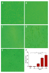
Cerebellar granule cells die when maintained in different of heroin (×200). DIV7 CGNs were then switched to medium containing 10 ug/ml, 40 ug/ml 80 ug/ml, 120 ug/ml heroin. Phase-contrast micrographs show neurons maintained in 10 ug/ml (B), 40 ug/ml (C), 80 ug/ml (D), 120 ug/ml (E) heroin for 24. Control cells (A) were maintained for 24 h in heroin-free medium. The percentages of death neurons under the treatments indicated were quantified (F). Scale bar=10 um. Data represent the means ±SEM of five independent experiments. * p<0.05,** p<0.01.
Figure 3
Figure 3
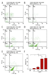
Crebellar granule cells apoptosis when maintained in different of heroin. DIV7 CGNs were then switched to medium containing 10 ug/ml (B), 40 ug/ml (C), 80 ug/ml (D), 120 ug/ml (E) heroin. Flow cytometry show neurons apoptosis maintained in different of heroin dosage for 24h. Control cells (A) were maintained for 24 h in heroin-free medium. The percentages of neurons apoptosis under the treatments indicated were quantified (F). Scale bar=10 um. Data represent the means ±SEM of three independent experiments. * p<0.05,** p<0.01.
Figure 4
Figure 4
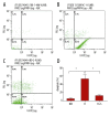
Cerebellar granule cells apoptosis when maintained in 120 ug/ml heroin (H) and SP600125+120ug/ml heroin (H+S). DIV7 CGNs were then switched to medium containing 120 ug/ml heroin (B), SP600125 +120 ug/ml heroin (C). Flow cytometry show neurons apoptosis maintained in 120ug/ml heroin and SP600125 +120 ug/ml heroin for 24 h. Control cells (A) were maintained for 24 h in normal medium. The percentages of neurons apoptosis under the treatments indicated were quantified (D). Scale bar=10 um. Data represent the means ±SEM of three independent experiments.*p <0.05.
Figure 5
Figure 5
JNK/c-Jun pathway regulates Cytc and ATF3 induction in heroin-induced neuronal apoptosis. (A) DIV7 CGNs were placed in media with or without different dosage of heroin for 24 h (a=normal medium, b=10 ug/ml, c=40 ug/ml, d=80 ug/ml, e=120 ug/ml), then neurons were assessed by PCR using the indicated the adopt of p-c-jun, cytc, ATF3 and β-actin primer (A1) and RT-PCR using the p-c-jun, cytc, ATF3 and β-actin were quantified (A2–A4). (B) DIV7 CGNs were placed in media with or without different dosage of heroin for 24 h (a=normal medium, b=10 ug/ml, c=40 ug/ml, d=80 ug/ml, e=120 ug/ml), then neurons were assessed by Western Blotting using the indicated the adopt of p-c-jun, cytc, ATF3 and β-tubulin protein (B2–B4). (C) DIV7 CGNs were transferred to media containing 120 ug/ml heroin in the absence or presence of 10 um SP600125.After 24 h, neurons were assessed by PCR using the indicated Semi-quantitative (C1) and RT-PCR using the p-c-jun, cytc and β-actin were quantified (C2–C4). (D) DIV7 CGNs were transferred to media containing 120 ug/ml heroin in the absence or presence of 10 um SP600125. After 24 h, neurons were assessed by Western Blotting using the p-c-jun, cytc and β-tubulin protein were quantified (D2–D4).* p<0.05, **p<0.01.

Similar articles

Cited by

References

    1. Jordan MT, Bryant SM, Aks SE, et al. A five-year review of the medical outcome of heroin body stuffers. J Emerg Med. 2009;36(3):250–56. - PubMed
    1. Nutt D, King LA, Saulsbury W, et al. Development of a rational scale to assess the harm of drugs of potential misuse. Lancet. 2007;369(9566):1047–53. - PubMed
    1. Di Chiara G, Bassareo V. Reward system and addiction: what dopamine does and doesn’t do. Curr Opin Pharmacol. 2007;7(1):69–76. - PubMed
    1. Büttner A, Mall G, Penning R, et al. The neuropathology of heroin abuse. Forensic Sci Int. 2000;113(1–3):435–42. - PubMed
    1. Vassilev LT, Vu BT, Graves B, et al. In vivo activation of the p53 pathway by small-molecule antagonists of MDM2. Science. 2004;303(5659):844–48. - PubMed

MeSH terms