. 2015 Apr 7;11(4):e1005145.
doi: 10.1371/journal.pgen.1005145. eCollection 2015 Apr.

Proteolysis of virulence regulator ToxR is associated with entry of Vibrio cholerae into a dormant state

Affiliations

Proteolysis of virulence regulator ToxR is associated with entry of Vibrio cholerae into a dormant state

Salvador Almagro-Moreno et al. PLoS Genet. .

Abstract

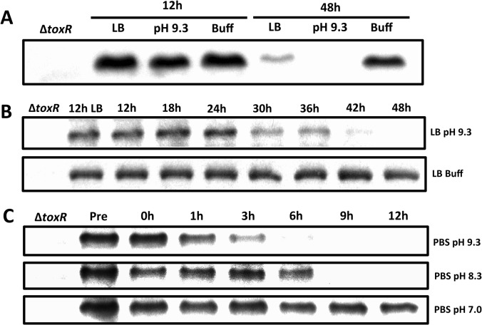
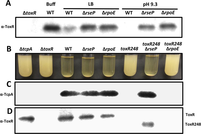
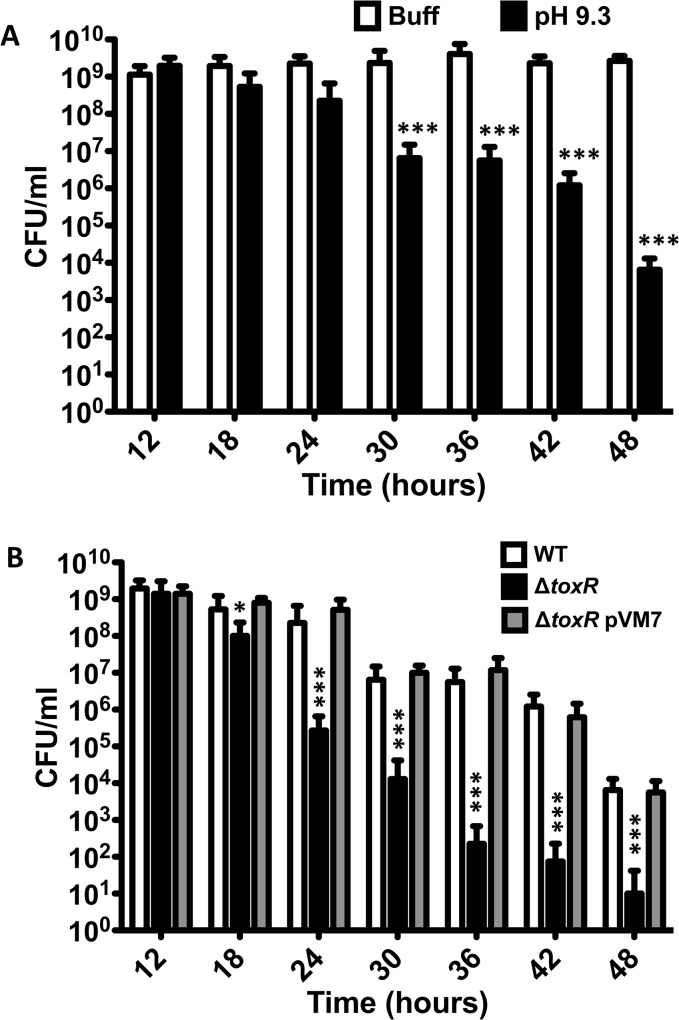
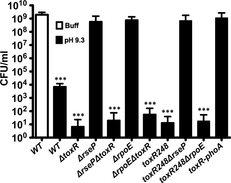
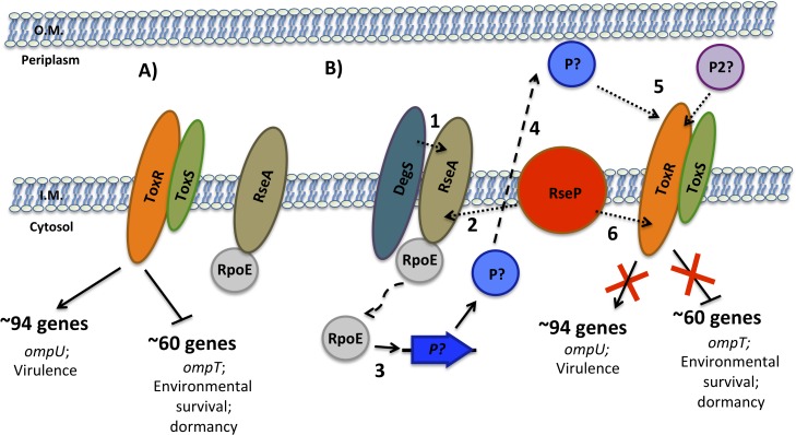
Vibrio cholerae O1 is a natural inhabitant of aquatic environments and causes the diarrheal disease, cholera. Two of its primary virulence regulators, TcpP and ToxR, are localized in the inner membrane. TcpP is encoded on the Vibrio Pathogenicity Island (VPI), a horizontally acquired mobile genetic element, and functions primarily in virulence gene regulation. TcpP has been shown to undergo regulated intramembrane proteolysis (RIP) in response to environmental conditions that are unfavorable for virulence gene expression. ToxR is encoded in the ancestral genome and is present in non-pathogenic strains of V. cholerae, indicating it has roles outside of the human host. In this study, we show that ToxR undergoes RIP in V. cholerae in response to nutrient limitation at alkaline pH, a condition that occurs during the stationary phase of growth. This process involves the site-2 protease RseP (YaeL), and is dependent upon the RpoE-mediated periplasmic stress response, as deletion mutants for the genes encoding these two proteins cannot proteolyze ToxR under nutrient limitation at alkaline pH. We determined that the loss of ToxR, genetically or by proteolysis, is associated with entry of V. cholerae into a dormant state in which the bacterium is normally found in the aquatic environment called viable but nonculturable (VBNC). Strains that can proteolyze ToxR, or do not encode it, lose culturability, experience a change in morphology associated with cells in VBNC, yet remain viable under nutrient limitation at alkaline pH. On the other hand, mutant strains that cannot proteolyze ToxR remain culturable and maintain the morphology of cells in an active state of growth. Overall, our findings provide a link between the proteolysis of a virulence regulator and the entry of a pathogen into an environmentally persistent state.


Conflict of interest statement

The authors have declared that no competing interests exist.

Figures

Fig 1
Fig 1. Proteolysis of ToxR during late stationary phase at alkaline pH.
(A) ToxR immunoblot of O395 wild-type or ΔtoxR grown for either 12 or 48 hours in LB starting pH 7.0 unbuffered (LB), LB starting pH 9.3 unbuffered (pH 9.3), or LB buffered to pH 7.0 with 100 mM HEPES (Buff). (B) ToxR immunoblots of O395 wild-type grown at different time points in LB starting pH 9.3 unbuffered (pH 9.3), or LB buffered to pH 7.0 with 100 mM HEPES (Buff). (C) ToxR immunoblots of O395 wild-type grown overnight in LB starting pH 7.0 unbuffered at 37°C, pelleted, and resuspended in phosphate buffered saline (PBS) at pH 7.0, pH 8.3, or pH 9.3 for 12 hours.
Fig 2
Fig 2. Proteolysis of ToxR is RseP and RpoE-dependent.
(A) ToxR immunoblot of cultures of O395 wild-type or ΔtoxR, ΔrseP or ΔrpoE grown for 48 hours in LB starting pH 7.0 unbuffered (LB), LB starting pH 9.3 unbuffered (pH 9.3), or LB buffered to pH 7.0 with 100 mM HEPES (Buff). (B) Autoagglutination of O395 ΔtcpA, ΔtoxR, wild-type (WT), ΔrseP, ΔrpoE, toxR248, toxR248ΔrseP and toxR248ΔrpoE grown under inducing conditions (LB starting pH 6.5, 30°C) for 15 hours. Autoagglutination can be visualized as a pellet at the bottom of the tube. (C) TcpA and (D) ToxR immunoblots of the cultures in (B).
Fig 3
Fig 3. V. cholerae shows reduced culturability over time at alkaline pH.
(A) CFU/ml of O395 wild-type strain grown at different time points in LB pH 7.0 with 100 mM HEPES (Buff), or LB starting pH 9.3 unbuffered (pH 9.3). The bars represent the mean of four independent experiments and the error bars indicate the standard deviation. Statistical comparisons were made using the student’s t-test and compare samples relative to 12h Buff. ***P < 0.0005. (B) CFU/ml of O395 wild-type (WT), ΔtoxR, or ΔtoxR pVM7 strains grown at different time points in LB starting pH 9.3 unbuffered. The bars represent the mean of four independent experiments and the error bars indicate the standard deviation. Statistical comparisons were made using the student’s t-test and compare samples relative to wild-type on LB pH 9.3 at that specific time point. *P < 0.05, ***P < 0.0005.
Fig 4
Fig 4. Reduction in culturability of V. cholerae depends on the loss of ToxR.
CFU/ml of O395 wild-type (WT) and mutant strains in LB pH 7.0 with 100 mM HEPES (Buff), or LB starting pH 9.3 unbuffered (pH 9.3) for 48 hours. The bars represent the mean of at least four independent experiments and the error bars indicate the standard deviation. Statistical comparisons were made using the student’s t-test and compare samples relative to wild-type 48h Buff. ***P < 0.0005.
Fig 5
Fig 5. Viability and morphology of V. cholerae after 48 hours at alkaline pH.
Fluorescent (F) and differential interference contrast (DIC) images of O395 wild-type grown in LB starting pH 7.0 unbuffered overnight and heat killed (O/N HK), LB starting pH 7.0 unbuffered overnight (O/N) as controls, 48 hours in LB buffered to pH 7.0 with 100 mM HEPES (48h Buff), and 48 hours in LB pH 9.3 unbuffered (48h pH 9.3).
Fig 6
Fig 6. Viability and morphology of V. cholerae mutants after 48 hours at alkaline pH.
Fluorescent (F) and differential interference contrast (DIC) images of O395 ΔtoxR, ΔrseP, ΔrsePΔtoxR, ΔrpoE or ΔrpoEΔtoxR, toxR248, toxR248ΔrseP, toxR248ΔrpoE, and toxR-phoA grown for 48 hours in LB starting pH 9.3 (unbuffered). The cells were observed after treatment with the LIVE/DEAD BacLight Bacterial Viability and Counting Kit. Viable and culturable cells appear green and elongated; viable but dormant cells appear green and round; dead cells appear red and round.
Fig 7
Fig 7. Effect of nutrient limitation in the stability of ToxR, culturability and morphology of Vibrio parahaemolyticus.
(A) ToxR immunoblot of V. parahaemolyticus RIMD2210633 wild-type or ΔtoxR grown for either 12 or 48 hours in LB starting pH 7.0 unbuffered (LB), or LB buffered to pH 7.0 with 100 mM HEPES (Buff) (B) Culturability of V. parahaemolyticus RIMD2210633 grown for 48 hours in LB starting pH 7.0 unbuffered (LB), or LB buffered to pH 7.0 with 100 mM HEPES (Buff). The bars represent the mean of four independent experiments and the error bars indicate the standard deviation. Statistical comparisons were made using the student’s t-test and compare samples relative to 48h Buff. ***P < 0.0005. (C) Fluorescent (F) and differential interference contrast (DIC) images of V. parahaemolyticus RIMD2210633 grown for 48 hours under conditions as in (B). The cells were observed after treatment with the LIVE/DEAD BacLight Bacterial Viability and Counting Kit. Viable and culturable cells appear green and elongated; viable but dormant cells appear green and round; dead cells appear red and round.
Fig 8
Fig 8. Sequential model for the RIP of ToxR during late stationary phase.
(A) In the early stages of colonization, when nutrients are abundant, ToxRS upregulate the expression of genes such as ompU important for growth under these conditions and downregulate the expression of genes such as ompT with roles in environmental survival. (B) During the late stages of colonization, as nutrients become depleted and the environment becomes alkalinized, ToxR is proteolyzed. This occurs due to activation of the σE pathway via sequential degradation of RseA by either DegS or another site-1 protease (1) and RseP (2), which releases RpoE to activate its regulated genes (3). One of these genes may encode a site-1 protease (P) that enters the periplasm (4) and cleaves a periplasmic portion of ToxR, however, at least a second protease/system (P2) appears to be necessary for the site-1 proteolytic event to occur (5). This event is followed by proteolysis of an inner-membrane site of ToxR by RseP (6), which then induces and prevents, respectively, expression of ToxR repressed and activated genes.

References

    1. Huq A, Small EB, West PA, Huq MI, Rahman R, et al. (1983) Ecological relationships between Vibrio cholerae and planktonic crustacean copepods. Appl Environ Microbiol 45: 275–283. - PMC - PubMed
    1. Tamplin ML, Gauzens AL, Huq A, Sack DA, Colwell RR (1990) Attachment of Vibrio cholerae serogroup O1 to zooplankton and phytoplankton of Bangladesh waters. Appl Environ Microbiol 56: 1977–1980. - PMC - PubMed
    1. Almagro-Moreno S, Taylor RK (2013) Cholera: Environmental Reservoirs and Impact on Disease Transmission. Microbiol Spectrum 1(2):OH-0003-2012. - PubMed
    1. Lutz C, Erken M, Noorian P, Sun S, McDougald D (2013) Environmental reservoirs and mechanisms of persistence of Vibrio cholerae . Front Microbiol 4: 375 10.3389/fmicb.2013.00375 - DOI - PMC - PubMed
    1. Taylor RK, Miller VL, Furlong DB, Mekalanos JJ (1987) Use of phoA gene fusions to identify a pilus colonization factor coordinately regulated with cholera toxin. Proc Natl Acad Sci USA 84: 2833–2837. - PMC - PubMed

Publication types

MeSH terms

LinkOut - more resources