. 2015 Mar 3:15:70.
doi: 10.1186/s12870-015-0455-z.

Parallel action of AtDRB2 and RdDM in the control of transposable element expression

Parallel action of AtDRB2 and RdDM in the control of transposable element expression

Marion Clavel et al. BMC Plant Biol. .

Abstract

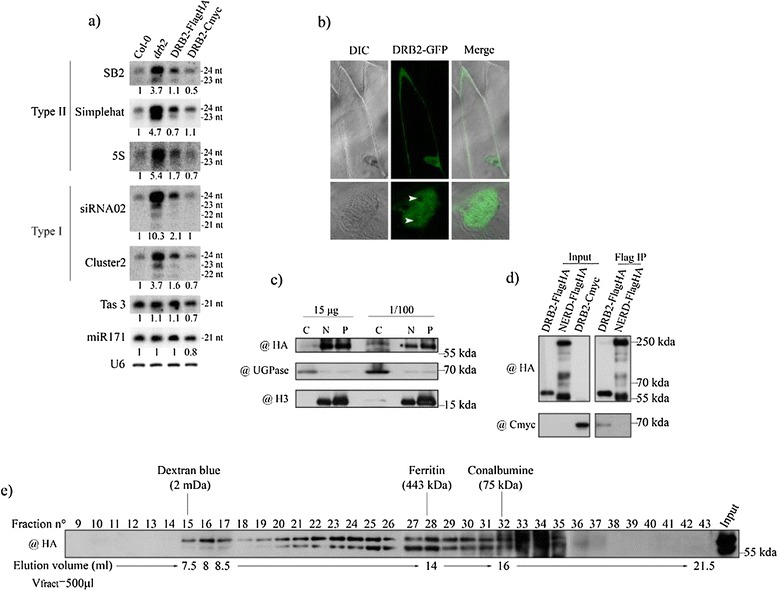
Background: In plants and animals, a large number of double-stranded RNA binding proteins (DRBs) have been shown to act as non-catalytic cofactors of DICERs and to participate in the biogenesis of small RNAs involved in RNA silencing. We have previously shown that the loss of Arabidopsis thaliana's DRB2 protein results in a significant increase in the population of RNA polymerase IV (p4) dependent siRNAs, which are involved in the RNA-directed DNA methylation (RdDM) process.

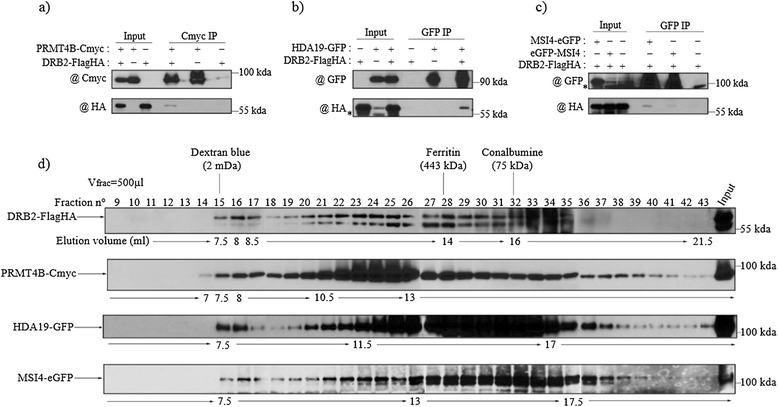
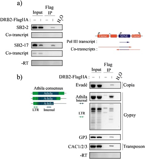
Results: Surprisingly, despite this observation, we show in this work that DRB2 is part of a high molecular weight complex that does not involve RdDM actors but several chromatin regulator proteins, such as MSI4, PRMT4B and HDA19. We show that DRB2 can bind transposable element (TE) transcripts in vivo but that drb2 mutants do not have a significant variation in TE DNA methylation.

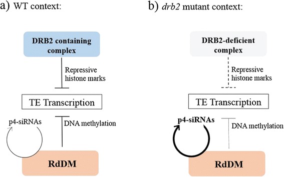
Conclusion: We propose that DRB2 is part of a repressive epigenetic regulator complex involved in a negative feedback loop, adjusting epigenetic state to transcription level at TE loci, in parallel of the RdDM pathway. Loss of DRB2 would mainly result in an increased production of TE transcripts, readily converted in p4-siRNAs by the RdDM machinery.


Figures

Figure 1
Figure 1
DRB2 is found predominantly in the nucleus and forms a high molecular weight complex as well as a homo interaction. (a) Level of small RNA accumulation in wild-type (Col-0), drb2-1 and two complementing lines. Values are normalized to U6 RNA and are expressed as a ratio relative to Col-0. For p4-siRNAs, only the 24-nt species were used for normalization. (b) Subcellular localization of DRB2-GFP in a heterologous system. GFP signal is observed both in the cytoplasm and the nucleus, but is absent from the nucleolus. (c) Subcellular localization of DRB2-FlagHA by cell fractionation and western blot. DRB2-FlagHA appears to be mainly nuclear. Extracts from each compartment were loaded in a SDS-PAGE either as a fixed protein quantity (first three lanes) or as 1/100th of the total extract (last three lanes). C stands for cytoplasm, N for nucleus and P for pellet. DRB2-FlagHA is revealed with a commercial HA antibody (@), UGPase is used as the cytosol quality control and H3 as the nuclear quality control. (d) Coimmunopurification of the DRB2-Cmyc protein from DRB2-FlagHA bound Flag magnetic beads. DRB2-FlagHA is able to bind DRB2-Cmyc, while NERD-FlagHA is not. DRB2-FlagHA and NERD-FlagHA are both revealed with a commercial HA antibody and the presence of DRB2-Cmyc in the DRB2-FlagHA eluate is revealed with a Cmyc commercial antibody. (e) Gel filtration on a Superose 6 column of DRB2-FlagHA crude extracts. The elution profile of DRB2-FlagHA shows that it is present in a high molecular weight complex of an approximate mass of 2 MDa as well as in the intermediate forms of lower mass of this complex. Fractions (500 μl) were analysed by western blot, and DRB2-FlagHA is revealed with HA antibody. Fraction numbers, sizing standards and corresponding volumes are indicated.
Figure 2
Figure 2
DRB2 interacts in planta with proteins involved in chromatin regulation. (a-c) Co-immunoprecipitations confirming the interaction between DRB2 and (a) PRMT4B, (b) HDA19, and (c) MSI4. For each experiment, F1 plants resulting from the cross between the two lines and harbouring both transgenes were used, while either the parental line or a F1 plant segregating only one of the transgenes were used as negative controls. Inputs and purified fractions were analysed by western blot. Background bands are indicated by an asterisk (*). (d) Gel filtration on a superose 6 column of DRB2-FlagHA, PRMT4B-Cmyc, HDA19-GFP and MSI4-eGFP crude extracts. Fractions (500 μl) were analysed by western blot and fraction numbers, sizing standards and corresponding volumes are indicated. In all cases, DRB2-FlagHA is revealed with a HA antibody, PRMT4B-Cmyc with a Cmyc antibody and HDA19-GFP, MSI4-eGFP, eGFP-MSI4 are revealed using a GFP antibody.
Figure 3
Figure 3
DRB2 is able to bind TE transcripts. (a) RNA Immunoprecipitation (RIP) from mixed floral tissues of SINE transcripts in DRB2-FlagHA x ddm1 plants and ddm1 plants included as a negative control. Total RNA is extracted following the IP, DNase treated and reverse transcribed. PCR amplification is performed with primers specific to one element, and a second set of primers specific to the putative co-transcript. Each time, a control reaction is performed with water instead of matrix cDNA (H2O), and each time, absence of contaminant genomic DNA is assessed by performing the same amplification with the non-reverse transcribed material (−RT). (b) Same RIP experiment performed on a diverse set of TEs, one Copia, two Gypsies and one CACTA. Primer sets used to amplify the Athila family are designed on a consensus sequence and can therefore amplify numerous genomic copies, both in the LTR and in the internal sequence. Evadé, GP3 are locus specific primers while CAC1/2/3 primers detect three different loci. The same control reactions are performed.
Figure 4
Figure 4
Proposed model for the action of the DRB2 containing complex, and the resulting situation in the drb2 mutant. (a) In wild type plants, both the RdDM and the DRB2 containing complex act independently to negatively regulate TE transcription. RdDM uses siRNA-mediated DNA methylation to induce silencing while targeting of the DRB2 complex to TE nascent transcript would directly result in an increase in chromatin repressive marks at these loci. (b) In a drb2 plant, targeting efficiency of the complex to nascent transcripts decreases leading to and increase in TE transcription. As no components of the RdDM are impaired, these transcripts are routed to DCL3/RDR2 for p4-siRNA biogenesis leading to the symptomatic over-accumulation of p4-siRNAs observed in the drb2 mutant without changing the steady state level of TE RNAs.

Similar articles

Cited by

References

    1. Tian B, Mathews MB. Phylogenetics and functions of the double-stranded RNA-binding motif: a genomic survey. Prog Nucleic Acid Res Mol Biol. 2003;74:123–58. doi: 10.1016/S0079-6603(03)01012-2. - DOI - PubMed
    1. Tian B, Bevilacqua PC, Diegelman-Parente A, Mathews MB. The double-stranded-RNA-binding motif: interference and much more. Nat Rev Mol Cell Biol. 2004;5(12):1013–23. doi: 10.1038/nrm1528. - DOI - PubMed
    1. Chang KY, Ramos A. The double-stranded RNA-binding motif, a versatile macromolecular docking platform. FEBS J. 2005;272(9):2109–17. doi: 10.1111/j.1742-4658.2005.04652.x. - DOI - PubMed
    1. Bevilacqua PC, Cech TR. Minor-groove recognition of double-stranded RNA by the double-stranded RNA-binding domain from the RNA-activated protein kinase PKR. Biochemistry. 1996;35(31):9983–94. doi: 10.1021/bi9607259. - DOI - PubMed
    1. Ryter JM, Schultz SC. Molecular basis of double-stranded RNA-protein interactions: structure of a dsRNA-binding domain complexed with dsRNA. EMBO J. 1998;17(24):7505–13. doi: 10.1093/emboj/17.24.7505. - DOI - PMC - PubMed

Publication types

LinkOut - more resources