Review
. 2015 Jan 30;17(1):12.
doi: 10.1186/s13058-015-0519-x.

Invasive lobular carcinoma of the breast: morphology, biomarkers and 'omics

Review

Invasive lobular carcinoma of the breast: morphology, biomarkers and 'omics

Amy E McCart Reed et al. Breast Cancer Res. .

Abstract

Invasive lobular carcinoma of the breast is the most common 'special' morphological subtype of breast cancer, comprising up to 15% of all cases. Tumours are generally of a good prognostic phenotype, being low histological grade and low mitotic index, hormone receptor positive and HER2, p53 and basal marker negative, and with a generally good response to endocrine therapy. Despite this, clinicians face countless challenges in the diagnosis and long-term management of patients, as they encounter a tumour that can be difficult to detect through screening, elicits a very invasive nature, a propensity for widespread metastatic colonisation and, consequently, in some studies a worse long-term poor outcome compared with invasive carcinoma of no special type. Here we review the morphological and molecular features that underpin the disparate biological and clinical characteristics of this fascinating tumour type.


Figures
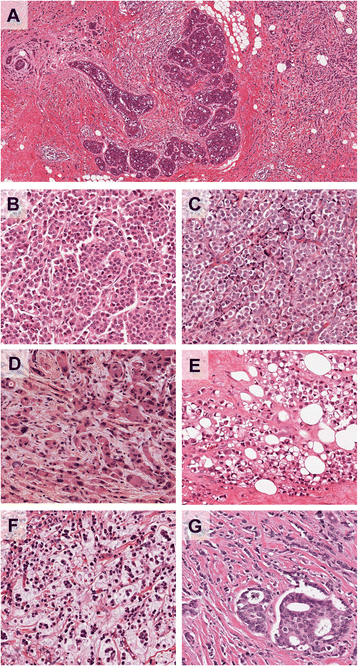
Figure 1
Figure 1
Morphological characteristics of invasive lobular carcinoma and its variants. (A) Low power view of a terminal duct lobular unit colonised by lobular carcinoma in situ. Classic invasive lobular carcinoma is seen diffusely infiltrating the whole specimen as single cells and single files of cells. The characteristic targetoid growth pattern is evident on the left-hand side (see also Figure 2). (B-G) Morphological variants of the classic type: (B) alveolar type, with globular aggregates of approximately 20 cells; (C) solid type with discohesive tumour cells growing in solid sheets; (D) a pleomorphic variant - note the pink, foamy cytoplasm typical of an apocrine phenotype and irregular nuclei; (E) pleomorphic invasive lobular carcinoma with prominent signet ring cells; (F) invasive lobular carcinoma showing mucinous/histiocytoid morphology; (G) mixed ductal-lobular carcinoma.
Figure 2
Figure 2
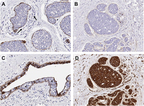
Immunohistochemical staining of E-cadherin and its binding complex in invasive lobular carcinoma. Lobular carcinoma in situ (LCIS) and invasive lobular carcinoma (ILC); note invasive neoplastic cells of ILC (arrows) growing in a targetoid fashion around the in situ component. (A) E-cadherin and (B) β-catenin staining is negative in both LCIS and ILC, although positive staining is observed in the myoepithelial cells surrounding the LCIS. (C) Pagetoid spread (asterisks) is also observed in this case, whereby the neoplastic cells (negative for E-cadherin) are growing and invading between the luminal and myoepithelial cells of a normal ductal structure). (D) In the absence of E-cadherin, there is a strong re-localisation of p120-catenin to the cytoplasm of neoplastic cells in LCIS and ILC.
Figure 3
Figure 3
Genomic architecture of invasive lobular carcinoma. (A) CIRCOS plot of an invasive lobular carcinoma (ILC) tumour profiled using the Illumina Omni 2.5 million SNP CNV array. Note the archetypal ILC changes, including chromosome 1q gain, 8p amplification, 11q13 amplification and 16q deletion. (B) Spectrum of somatic mutations across the E-cadherin coding region in the cBioPortal ILC data set [30,31]. Note the cadherin prodomain in green and the cadherin cytoplasmic domain in blue; missense mutations in green and nonsense mutations in red. (C) Oncoprint depicting the frequency of somatic mutations in key, recurrently altered cancer genes (CDH1, TP53, PIK3CA, ERBB2) affecting 75% of the 155 ILCs in The Cancer Genome Atlas cohort [30,31]. Percentages are numbers of tumours exhibiting an alteration in the specified gene.

References

    1. Ferlicot S, Vincent-Salomon A, Medioni J, Genin P, Rosty C, Sigal-Zafrani B, Freneaux P, Jouve M, Thiery JP, Sastre-Garau X. Wide metastatic spreading in infiltrating lobular carcinoma of the breast. Eur J Cancer. 2004;40:336–41. doi: 10.1016/j.ejca.2003.08.007. - DOI - PubMed
    1. Rakha EA, El-Sayed ME, Powe DG, Green AR, Habashy H, Grainge MJ, Robertson JF, Blamey R, Gee J, Nicholson RI, Lee AH, Ellis IO. Invasive lobular carcinoma of the breast: response to hormonal therapy and outcomes. Eur J Cancer. 2008;44:73–83. doi: 10.1016/j.ejca.2007.10.009. - DOI - PubMed
    1. Pestalozzi BC, Zahrieh D, Mallon E, Gusterson BA, Price KN, Gelber RD, Holmberg SB, Lindtner J, Snyder R, Thurlimann B, Murray E, Viale G, Castiglione-Gertsch M, Coates AS, Goldhirsch A, International Breast Cancer Study Group Distinct clinical and prognostic features of infiltrating lobular carcinoma of the breast: combined results of 15 International Breast Cancer Study Group clinical trials. J Clin Oncol. 2008;26:3006–14. doi: 10.1200/JCO.2007.14.9336. - DOI - PubMed
    1. Lakhani SR, Ellis IO, Schnitt SJ, Tan PH, van de Vijver MJ. WHO Classification of Tumours of the Breast. 4. Lyon: IARC; 2012.
    1. Haagensen CD, Lane N, Lattes R, Bodian C. Lobular neoplasia (so-called lobular carcinoma in situ) of the breast. Cancer. 1978;42:737–69. doi: 10.1002/1097-0142(197808)42:2<737::AID-CNCR2820420247>3.0.CO;2-T. - DOI - PubMed

Publication types

MeSH terms