. 2015 Apr 7:6:6510.
doi: 10.1038/ncomms7510.

Symportin 1 chaperones 5S RNP assembly during ribosome biogenesis by occupying an essential rRNA-binding site

Affiliations

Symportin 1 chaperones 5S RNP assembly during ribosome biogenesis by occupying an essential rRNA-binding site

Fabiola R Calviño et al. Nat Commun. .

Abstract

During 60S biogenesis, mature 5S RNP consisting of 5S RNA, RpL5 and RpL11, assembles into a pre-60S particle, where docking relies on RpL11 interacting with helix 84 (H84) of the 25S RNA. How 5S RNP is assembled for recruitment into the pre-60S is not known. Here we report the crystal structure of a ternary symportin Syo1-RpL5-N-RpL11 complex and provide biochemical and structural insights into 5S RNP assembly. Syo1 guards the 25S RNA-binding surface on RpL11 and competes with H84 for binding. Pull-down experiments show that H84 releases RpL11 from the ternary complex, but not in the presence of 5S RNA. Crosslinking mass spectrometry visualizes structural rearrangements on incorporation of 5S RNA into the Syo1-RpL5-RpL11 complex supporting the formation of a pre-5S RNP. Our data underline the dual role of Syo1 in ribosomal protein transport and as an assembly platform for 5S RNP.


Figures
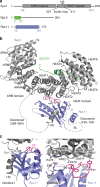
Figure 1
Figure 1. Structure of the Syo1–RpL5-N–RpL11 complex.
(a) Domain architecture of Syo1 (75 kDa), RpL5 (35 kDa) and RpL11 (25 kDa). Domains present in the crystal structure are given by residue numbers and are highlighted in colour. BL, basic loop; HS, helical segment from the acidic loop. (b) Overall structure of Syo1 (grey) in complex with RpL5-N (green) and RpL11 (blue). Disordered regions are shown as dashed lines, with residue numbers. N- and C-termini are indicated. (c) Close up of Syo1 (grey) and RpL11 (blue) interactions (interface I and II). Loop L3 is highlighted in red; HEAT repeats 1 and 2 are labelled.
Figure 2
Figure 2. Syo1 mimics 25S RNA (helix 84) binding to RpL11.
(a) RpL11 interaction with Syo1 (this study; middle), with helix 84 (as part of the ribosome; left16) are compared by superposition of both complexes (right; rotated by 90°). Syo1-HS and H84 share the binding site on RpL11 and read out its fold. (b) Binding of 5S RNA was tested by EMSAs. The titrations are shown for Syo1–RpL5–RpL11 (panel 1), Syo1–RpL5–RpL11ΔBL (panel 2), Syo1–RpL5ΔC–RpL11ΔBL (panel 3), Syo1–RpL5-N–RpL11 (panel 4), Syo1–RpL11 (panel 5) and Syo1–RpL11ΔBL complexes (panel 6). ‘RNA' lanes are free of protein to provide a base line for the shift. The protein/RNA ratio is given at the top. (c) Pull-down analysis of H84 interaction with the Syo1–RpL5–RpL11 complex (all proteins are full length). A GST-RpL11 variant was used to immobilize the complex on a GSTrap HP column (lane I). Washing with H84 releases Syo1–RpL5 (lane W) from GST-RpL11 (lane E).
Figure 3
Figure 3. Syo1 serves as a platform for 5S RNP assembly.
(a) Inter- (orange) and intraprotein (blue lines) crosslinks detected on the Syo1 complex in the absence (left) or presence (right) of the 5S RNA. Quantified crosslinks are shown in Table 2 and Supplementary Fig. 9. (b) Model for the role of Syo1 in 5S RNP assembly (left and middle panel). H84 is recruited into the tetrameric pre-5S RNP (right panel, top) by interaction with RpL11 as used in 5S RNP docking to the pre-60S particle (right panel, bottom). Parts of the proteins absent or disordered in the ternary complex are represented by dotted lines. Hinge points (represented as thick circles) in the proteins allow for rearrangements (indicated by arrows). Syo1 (grey), RpL5 (green), RpL11 (blue), 5S RNA (red) and helix 84 (H84, orange). The model is based on the ternary complex (this study) and the 5S RNP as part of the yeast ribosome (PDB codes: 3U5E, 3U5D). H84 interacts with the 5S RNP in the pre-60S and in the mature ribosome in the same manner.

References

    1. Jenner L. et al. Crystal structure of the 80S yeast ribosome. Curr. Opin. Struct. Biol. 22, 759–767 (2012) . - PubMed
    1. Kressler D., Hurt E. & Bassler J. Driving ribosome assembly. Biochim. Biophys. Acta 1803, 673–683 (2010) . - PubMed
    1. Tschochner H. & Hurt E. Pre-ribosomes on the road from the nucleolus to the cytoplasm. Trends Cell Biol. 13, 255–263 (2003) . - PubMed
    1. Venema J. & Tollervey D. Ribosome synthesis in Saccharomyces cerevisiae. Annu. Rev. Genet. 33, 261–311 (1999) . - PubMed
    1. Bange G., Murat G., Sinning I., Hurt E. & Kressler D. New twist to nuclear import: when two travel together. Commun. Integr. Biol. 6, e24792 (2013) . - PMC - PubMed

Publication types

Associated data

LinkOut - more resources