. 2015 Apr 7;10(4):e0123717.
doi: 10.1371/journal.pone.0123717. eCollection 2015.

Tissue factor-expressing tumor cells can bind to immobilized recombinant tissue factor pathway inhibitor under static and shear conditions in vitro

Affiliations

Tissue factor-expressing tumor cells can bind to immobilized recombinant tissue factor pathway inhibitor under static and shear conditions in vitro

Sara P Y Che et al. PLoS One. .

Abstract

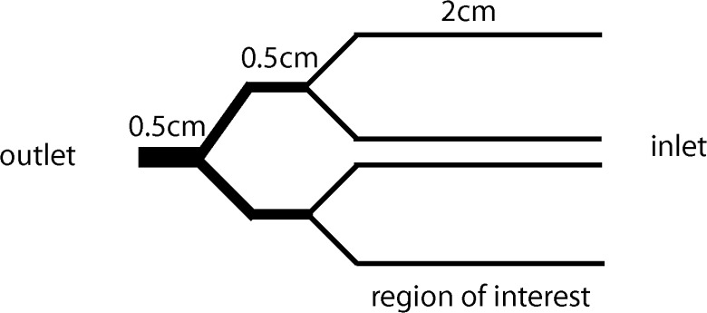
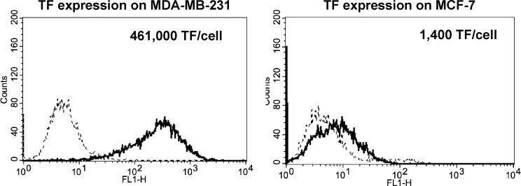
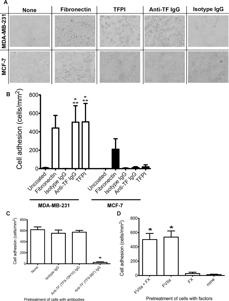
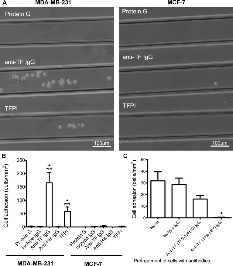
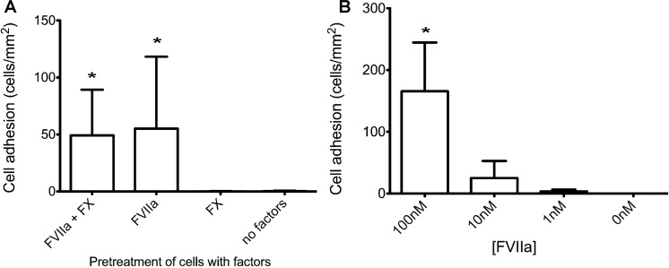
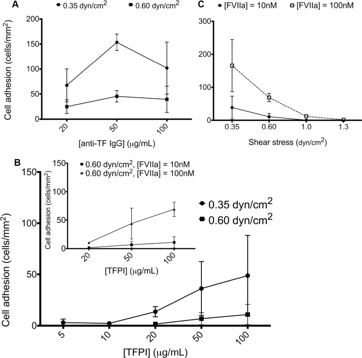
Mammary tumors and malignant breast cancer cell lines over-express the coagulation factor, tissue factor (TF). High expression of TF is associated with a poor prognosis in breast cancer. Tissue factor pathway inhibitor (TFPI), the endogenous inhibitor of TF, is constitutively expressed on the endothelium. We hypothesized that TF-expressing tumor cells can bind to immobilized recombinant TFPI, leading to arrest of the tumor cells under shear in vitro. We evaluated the adhesion of breast cancer cells to immobilized TFPI under static and shear conditions (0.35 - 1.3 dyn/cm2). We found that high-TF-expressing breast cancer cells, MDA-MB-231 (with a TF density of 460,000/cell), but not low TF-expressing MCF-7 (with a TF density of 1,400/cell), adhered to recombinant TFPI, under static and shear conditions. Adhesion of MDA-MB-231 cells to TFPI required activated factor VII (FVIIa), but not FX, and was inhibited by a factor VIIa-blocking anti-TF antibody. Under shear, adhesion to TFPI was dependent on the TFPI-coating concentration, FVIIa concentration and shear stress, with no observed adhesion at shear stresses greater than 1.0 dyn/cm2. This is the first study showing that TF-expressing tumor cells can be captured by immobilized TFPI, a ligand constitutively expressed on the endothelium, under low shear in vitro. Based on our results, we hypothesize that TFPI could be a novel ligand mediating the arrest of TF-expressing tumor cells in high TFPI-expressing vessels under conditions of low shear during metastasis.


Conflict of interest statement

Competing Interests: The authors have declared that no competing interests exist.

Figures

Fig 1
Fig 1. Schematic of microfluidic channel.
The microfluidic channel consisted of four branches (120x120μm), which allowed for four simultaneous experiments under different coating conditions or cell treatments. The indicated region of interest (along the length of the 4 branches) is where adherent cells are quantified. Cell suspensions were introduced at the inlet and the outlet was connected to a syringe pump.
Fig 2
Fig 2. TF surface expression and density on breast cancer cells.
Representative fluorescence histograms of TF expression on MDA-MB-231 and MCF-7 cells. Cells (5x10⁵) were incubated with a monoclonal antibody against TF (TF9-5B7, 80μg/mL), followed by an Alexa-488-conjugated secondary antibody (10μg/mL). Fluorescence was detected (bold line) using flow cytometry with isotype IgG as a control (dotted line). Tissue factor was strongly expressed on MDA-MB-231, but little expression was found on MCF-7 (n = 3). The surface ligand density is also shown for each cell line.
Fig 3
Fig 3. Static adhesion of tumor cells to protein-immobilized poly-dimethylsiloxane wells.
MDA-MB-231 and MCF-7 (5x10⁴ cells) were incubated for 1 hour at 37°C in PDMS wells immobilized with TFPI (50μg/ml), using anti-TF IgG (TF9-5B7, 50μg/ml) as a positive control for TF-specific adhesion, fibronectin (10μg/ml) as a positive control for integrin adhesion, and uncoated or isotype IgG (50μg/ml) coated wells as negative controls. For TFPI-treated wells, cells were pretreated with 10nM FVIIa and 10nM FX for 10 minutes prior to addition to the wells. A. Representative bright field images of adherent cells on the different coatings. More MDA-MB-231 bound to TFPI- and anti-TF antibody-coated wells than MCF-7 cells. Both cell lines bound to fibronectin-coated wells. B. Adherent cells were counted and normalized by the area of the counted region (mean ± standard deviation). Significantly more MDA-MB-231 cells bound to TFPI- and anti-TF IgG-coated wells than MCF-7 (* p < 0.05, n = 6 for TFPI, n = 3 for anti-TF IgG). Significantly more MDA-MB-231 bound to TFPI- and anti-TF IgG-coated wells than uncoated or isotype IgG-coated wells (** p<0.05). C. MDA-MB-231 cells were pretreated with 50μg/ml anti-TF IgG (TF9-5B7 which blocks FVIIa binding to TF, and TF9-10H10 which does not block FVIIa binding to TF). The positive control had no antibody pretreatment, and isotype IgG pretreatment (50μg/ml) was used as a negative control. Blocking FVIIa binding to TF with the TF9-5B7 antibody significantly decreased adhesion to TFPI-coated wells compared to controls (* p < 0.05, n = 3). D. MDA-MB-231 cells were treated with different combinations of FVIIa (10nM) and FX (10nM) before incubation with TFPI-coated wells. Adhesion to TFPI was significantly decreased only when FVIIa was absent (* p < 0.05, n = 3).
Fig 4
Fig 4. Adhesion of tumor cells to protein-immobilized microfluidic channels under low shear (0.35dyn/cm2).
Microfluidic channels were incubated with Protein G (100μg/ml), then anti-TF IgG (100μg/ml), or an anti-His antibody (100μg/ml) followed by TFPI (100μg/ml). Isotype IgG (100μg/ml) and anti-His IgG (100μg/ml) antibodies were used as negative control for anti-TF IgG and TFPI respectively. Tumor cells (1x10⁶cells/mL, pre-treated with 10nM FVIIa and 10nM FX for TFPI-coated channels) were introduced into the channels at 0.35dyn/cm2 for 30 minutes, and non-specifically adhered cells were removed at 2.0dyn/cm2. The entire channel was imaged to quantify the number of adherent cells. A. Representative bright field images of adherent tumor cells on channels immobilized with Protein G (negative control), anti-TF IgG and TFPI showing that more MDA-MB-231 than MCF-7 cells were bound to both anti-TF IgG- and TFPI-coated channels. B. The number of adherent cells was counted and normalized by the channel area. MDA-MB-231 showed significantly higher adhesion to TFPI- and anti-TF IgG-coated channels than MCF-7 (* p < 0.05, n = 4 for anti-TF IgG, n = 3 for TFPI). Significantly more MDA-MB-231 bound to TFPI- and anti-TF IgG-coated channels than negative controls (** p<0.05). C. MDA-MB-231 cells were pretreated with 50μg/ml anti-TF IgG (TF9-5B7 which blocks FVIIa binding to TF, or TF9-10H10 which does not block FVIIa binding to TF). The positive control had no antibody pretreatment, and isotype IgG pretreatment (50μg/ml) was used as a negative control. Blocking FVIIa binding to TF with TF9-5B7 antibody significantly decreased adhesion to TFPI-coated channels (* p < 0.05, n = 4). The observed decrease in MDA-MB-231 adhesion with the TF9-10H10 antibody, albeit not significant with this stringent statistical test, could be due to steric hindrance of TFPI binding to the TF/FVIIa/FXa complex on the tumor cells.
Fig 5
Fig 5. Effect of FVIIa and FX in adhesion of MDA-MB-231 to TFPI-immobilized channels (0.35dyn/cm2).
A. MDA-MB-231 cells were treated with different combinations of FVIIa (10nM) and FX (10nM) before introduction into TFPI-immobilized channels. Adhesion of MDA-MB-231 to immobilized TFPI in microfluidic channels was abolished when FVIIa was absent (* p<0.05, n = 6). B. MDA-MB-231 cells were treated with different concentrations of FVIIa (0–100nM) prior to perfusion with TFPI-immobilized channels. Increasing the concentration of FVIIa to 100nM significantly increased adhesion of MDA-MB-231 to immobilized TFPI in microfluidic channels (* p < 0.05, n = 3).
Fig 6
Fig 6. Effect of shear, TFPI-coating concentration and FVIIa concentration in MDA-MB-231 adhesion to protein-immobilized channels under shear.
A. Microfluidic channels were immobilized with different concentrations of anti-TF IgG antibody (20–100μg/mL), and MDA-MB-231 cells were introduced at a shear of 0.35 and 0.60dyn/cm2. Adhesion of MDA-MB-231 cells to anti-TF IgG antibody reached a plateau at 50μg/mL at both shear stresses (n = 3). B. Microfluidic channels were immobilized with different concentrations of TFPI (5–100μg/mL), and MDA-MB-231 cells (pretreated with 10nM FVIIa and FX) were introduced at a shear of 0.35 and 0.60dyn/cm2. The adhesion of MDA-MB-231 increased with increasing TFPI concentration (n = 3). Inset. When FVIIa concentration was increased from 10nM to 100nM at a shear of 0.60dyn/cm2, the adhesion of MDA-MB-231 to TFPI-coated channels increased (n = 3). C. Microfluidic channels were immobilized with 100μg/mL TFPI, and MDA-MB-231 cells (pretreated with 10nM or 100nM FVIIa, and 10nM FX) were introduced at a range of shear stresses (0.35–1.3dyn/cm2). The adhesion of MDA-MB-231 decreased with increasing shear. Increasing the concentration of FVIIa from 10nM to 100nM increased adhesion of MDA-MB-231 to immobilized TFPI at 0.35 and 0.60dyn/cm2. A few tumor cells bound at 1.3dyn/cm2 with the higher, but not the lower, FVIIa concentration (n = 3).

References

    1. Bluff JE, Brown NJ, Reed MWR, Staton CA (2008) Tissue factor, angiogenesis and tumour progression. BCR 10: 204–213. 10.1186/bcr1871 - DOI - PMC - PubMed
    1. Rak J, Milsom C, Magnus N, Yu J (2009) Tissue factor in tumour progression. Best Pract Res Clin Haemato 22: 71–83. 10.1016/j.beha.2008.12.008 - DOI - PubMed
    1. Van den Berg YW, Osanto S, Reitsma PH, Versteeg HH (2012) The relationship between tissue factor and cancer progression: insights from bench and bedside. Blood 119: 924–932. 10.1182/blood-2011-06-317685 - DOI - PubMed
    1. Manly DA, Boles J, Mackman N (2011) Role of tissue factor in venous thrombosis. Annu Rev Physiol 73: 515–525. 10.1146/annurev-physiol-042210-121137 - DOI - PMC - PubMed
    1. Rickles FR, Patierno S, Fernandez PM (2003) Tissue Factor, thrombin, and cancer. Chest 124: 58S–68S. 10.1378/chest.124.3 - DOI - PubMed

Publication types