. 2015 Feb 13:14:22.
doi: 10.1186/s12933-015-0183-6.

Activation of TRPV1 channel by dietary capsaicin improves visceral fat remodeling through connexin43-mediated Ca2+ influx

Activation of TRPV1 channel by dietary capsaicin improves visceral fat remodeling through connexin43-mediated Ca2+ influx

Jian Chen et al. Cardiovasc Diabetol. .

Abstract

Background: The prevalence of obesity has dramatically increased worldwide and has attracted rising attention, but the mechanism is still unclear. Previous studies revealed that transient receptor potential vanilloid 1 (TRPV1) channels take part in weight loss by enhancing intracellular Ca2+ levels. However, the potential mechanism of the effect of dietary capsaicin on obesity is not completely understood. Ca2+ transfer induced by connexin43 (Cx43) molecules between coupled cells takes part in adipocyte differentiation. Whether TRPV1-evoked alterations in Cx43-mediated adipocyte-to-adipocyte communication play a role in obesity is unknown.

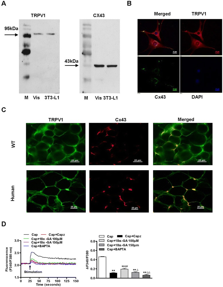
Materials and methods: We investigated whether Cx43 participated in TRPV1-mediated adipocyte lipolysis in cultured 3T3-L1 preadipocytes and visceral adipose tissues from humans and wild-type (WT) and TRPV1-deficient (TRPV1-/-) mice.

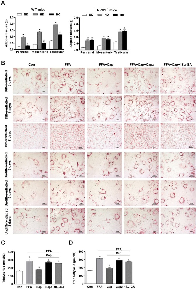
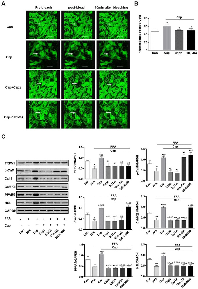
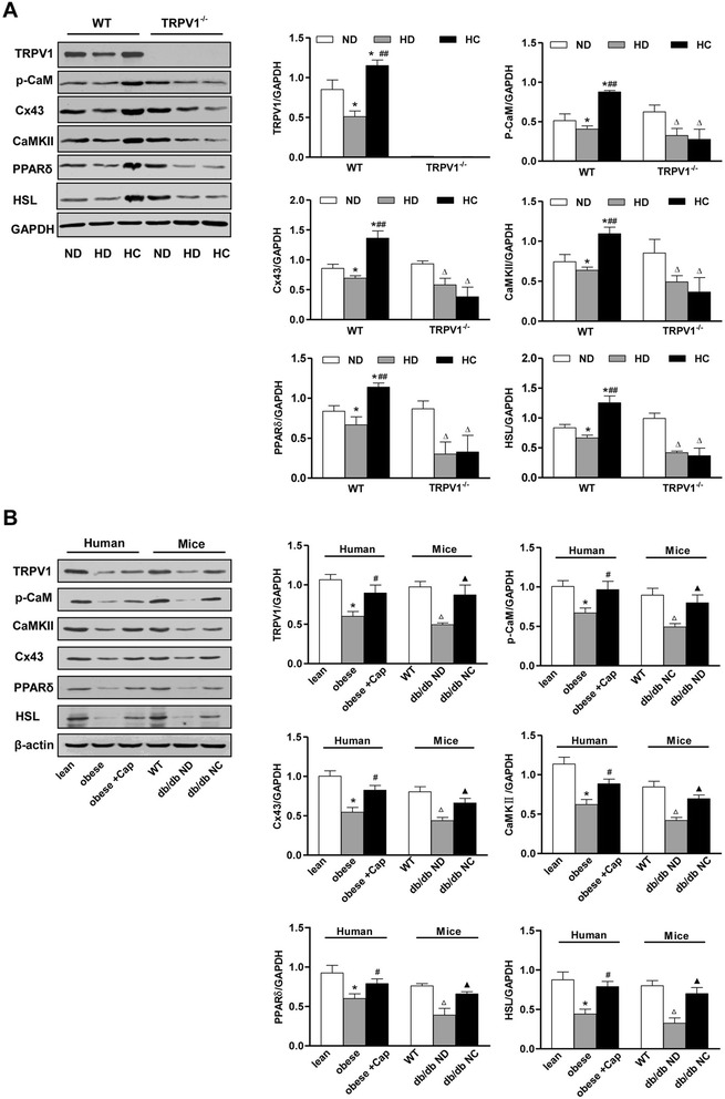
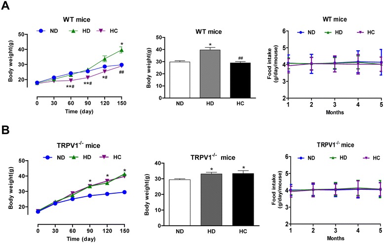
Results: TRPV1 and Cx43 co-expressed in mesenteric adipose tissue. TRPV1 activation by capsaicin increased the influx of Ca2+ in 3T3-L1 preadipocytes and promoted cell lipolysis, as shown by Oil-red O staining. These effects were deficient when capsazepine, a TRPV1 antagonist, and 18 alpha-glycyrrhetinic acid (18α-GA), a gap-junction inhibitor, were administered. Long-term chronic dietary capsaicin reduced the weights of perirenal, mesenteric and testicular adipose tissues in WT mice fed a high-fat diet. Capsaicin increased the expression levels of p-CaM, Cx43, CaMKII, PPARδ and HSL in mesenteric adipose tissues from WT mice fed a high-fat diet, db/db mice, as well as obese humans, but these effects of capsaicin were absent in TRPV1-/- mice. Long-term chronic dietary capsaicin decreased the body weights and serum lipids of WT mice, but not TRPV1-/- mice, fed a high-fat diet.

Conclusion: This study demonstrated that capsaicin activation of TRPV1-evoked increased Ca2+ influx in Cx43-mediated adipocyte-to-adipocyte communication promotes lipolysis in both vitro and vivo. TRPV1 activation by dietary capsaicin improves visceral fat remodeling through the up-regulation of Cx43.


Figures

Figure 1
Figure 1
Cx43 affects TRPV1-mediated Ca 2+ influx in adipocytes. A. TRPV1 and Cx43 were detected in mesenteric adipose tissue (Vis) and 3T3-L1 preadipocytes (3T3-L1). M indicates the marker. B and C. Immunofluorescence demonstrated specific staining for co-expressed TRPV1 and Cx43 in the cell-cell connecting portions in primary cultured human visceral adipocytes B and visceral adipose tissues from both wild-type mice (WT) and humans C. The green fluorescence indicates Cx43. The red fluorescence indicates TRPV1. Nuclei in all groups were stained in blue with DAPI. The images were collected using a Nikon TE2000-U inverted fluorescence microscope and are representative of 3 separate experiments. The scale bar indicates 25 μm. D. Representative curves (left panel) show [Ca2+]i changes in 3T3-L1 preadipocytes acutely stimulated with capsaicin (Cap, 1 μmol/L), capsaicin with the TRPV1 antagonist capsazepine (Cap + Capz, 1 μmol/L), the Cx43 inhibitor, 18α-glycyrrhetinic acid (Cap + 18α-GA, 100 or 150 μmol/L), or the intracellular Ca2+ chelator, BAPTA-AM (10 μmol/L). The summary data (right panel) show the maximal stimulated changes of [Ca2+]i (25-150 s) from the baseline (0-25 s). Values were expressed as the mean ± SEM; n = 4 per group. **P < 0.01 vs. Cap; ##P < 0.01 vs. Cap + Capz; ΔP < 0.05, ΔΔP < 0.01 vs. Cap + 18α-GA 100 μM.
Figure 2
Figure 2
TRPV1 activation promotes lipolysis both in mesenteric adipose tissue and 3T3-L1 preadipocytes. A. The weights of mesenteric, perirenal and testicular adipose tissues from WT and TRPV1−/− mice fed a normal diet (ND), a high-fat diet (HD), or a high-fat plus capsaicin diet (HC). *P < 0.05 vs. ND, #P < 0.05 vs. HD. Values are expressed as the mean ± SEM, n = 6. B. Lipid droplets were visualized by Oil red O staining of 3T3-L1 preadipocytes on days 3, 5, and 8, with or without differentiation. The cells were exposed to a mixture of 1 mmol/L long-chain free fatty acids (FFA) in the presence of 1 μmol/L capsaicin (FFA + Cap), capsaicin plus 1 μmol/L capsazepine (FFA + Cap + Capz) or 150 μmol/L 18α-GA (FFA + Cap + 18α-GA) for 24 h. Red spots indicate lipid droplets in the cells. Images are representative of 3 separate experiments. The scale bar indicates 10 μm. C and D. Triglyceride and free fatty acid levels of 3T3-L1 preadipocytes after treatment with free fatty acids (FFA), FFA + capsaicin (Cap), FFA + capsaicin + capsazepine (Capz) or FFA + capsaicin + 18α-GA (18α-GA, 150 μmol/L) for 24 h. *P < 0.05 vs Con, #P < 0.05 vs FFA, ΔP < 0.05 vs Cap. Values are expressed as the mean ± SEM; n = 3 per group.
Figure 3
Figure 3
TRPV1 promotes lipolysis of 3T3-L1 preadipocytes through the regulation of Cx43-mediated intracellular calcium levels. A. Images indicate the Fluorescence Recovery After Photobleaching (FRAP), after treatment with capsaicin (Cap), capsaicin + capsazepine (Capz) or capsaicin + 18α-GA (18α-GA) for 24 h, used to measure the dynamics of Cx43 mobility. B The ratio of fluorescence recovery inside the bleached regions. *P < 0.05 vs Con, #P < 0.05 vs Cap. The values were expressed as the mean ± SEM from 3 separate experiments. C. Immunoblots of TRPV1, p-CaM, Cx43, CaMKII, PPARδ, and HSL in 3T3-L1 preadipocytes treated with free fatty acids (FFA), FFA + capsaicin (Cap), FFA + capsaicin + capsazepine (Capz), FFA + capsaicin + EGTA (EGTA), FFA + capsaicin + 18α-GA (18α-GA) or FFA + capsaicin + GSK0660 (GSK0660, PPARδ inhibitor, 10 μmol/L) for 24 h. *P < 0.05, **P < 0.01 vs Con; #P < 0.05, ##P < 0.01vs FFA; ΔP < 0.05, ΔΔP < 0.01 vs Cap. The densitometric values of protein expression levels were all normalized to GAPDH. Values are expressed as the mean ± SEM; n = 3 per group.
Figure 4
Figure 4
TRPV1 activation by dietary capsaicin increases Cx43-mediated lipolysis of mesenteric adipose tissues in mice and humans. A. Immunoblots of TRPV1, p-CaM, Cx43, CaMKII, PPARδ, and HSL in mesenteric adipose tissues from WT and TRPV1-/- mice fed a ND, HD and HC. *P < 0.05 vs. WT ND, ##P < 0.01 vs. WT HD, ΔP < 0.05 vs. TRPV1-/- ND. B. Immunoblots of TRPV1, p-CaM, Cx43, CaMKII, PPARδ, and HSL in visceral adipose tissues from humans and WT and db/db mice. Obese + Cap indicates that visceral adipose tissues from obese humans were treated with capsaicin for 24 hours. Db/db mice were fed a standard laboratory chow (db/db ND) or standard laboratory chow plus 0.01% capsaicin (db/db NC). *P < 0.05 vs lean people, #P < 0.05 vs obese people, ΔP < 0.05 vs WT, P < 0.05 vs db/db ND. The densitometric values of protein expression levels were all normalized to GAPDH or β-actin. Values are expressed as the mean ± SEM for 3 mice or people.
Figure 5
Figure 5
TRPV1 activation by dietary capsaicin improves high-fat diet-induced obesity. A and B. Body weight gain and food intake of WT A and TRPV1−/− B mice fed with ND, HD and HC. The values are expressed as the mean ± SEM for 15 mice. *P < 0.05, **P < 0.01 vs. ND, ##P < 0.01 vs. HD.

References

    1. Gutierrez-Aguilar R, Kim DH, Woods SC, Seeley RJ. Expression of new loci associated with obesity in diet-induced obese rats: from genetics to physiology. Obesity (Silver Spring) 2012;20(2):306–12. doi: 10.1038/oby.2011.236. - DOI - PubMed
    1. Després JP. Abdominal obesity and cardiovascular disease: is inflammation the missing link? Can J Cardiol. 2012;28(6):642–52. doi: 10.1016/j.cjca.2012.06.004. - DOI - PubMed
    1. de Souza RJ, Bray GA, Carey VJ, Hall KD, LeBoff MS, Loria CM, et al. Effects of 4 weight-loss diets differing in fat, protein, and carbohydrate on fat mass, lean mass, visceral adipose tissue, and hepatic fat: results from the POUNDS LOST trial. Am J Clin Nutr. 2012;95(3):614–25. doi: 10.3945/ajcn.111.026328. - DOI - PMC - PubMed
    1. Despres JP, Lemieux I. Abdominal obesity and metabolic syndrome. Nature. 2006;444(7121):881–7. doi: 10.1038/nature05488. - DOI - PubMed
    1. Fox CS, Massaro JM, Hoffmann U, Pou KM, Maurovich-Horvat P, Liu CY, et al. Abdominal visceral and subcutaneous adipose tissue compartments: association with metabolic risk factors in the Framingham Heart Study. Circulation. 2007;116(1):39–48. doi: 10.1161/CIRCULATIONAHA.106.675355. - DOI - PubMed

Publication types