. 2015 Apr;71(Pt 4):986-95.
doi: 10.1107/S1399004715002527. Epub 2015 Mar 27.

Structural and functional characterization of TesB from Yersinia pestis reveals a unique octameric arrangement of hotdog domains

Affiliations

Structural and functional characterization of TesB from Yersinia pestis reveals a unique octameric arrangement of hotdog domains

C M D Swarbrick et al. Acta Crystallogr D Biol Crystallogr. 2015 Apr.

Abstract

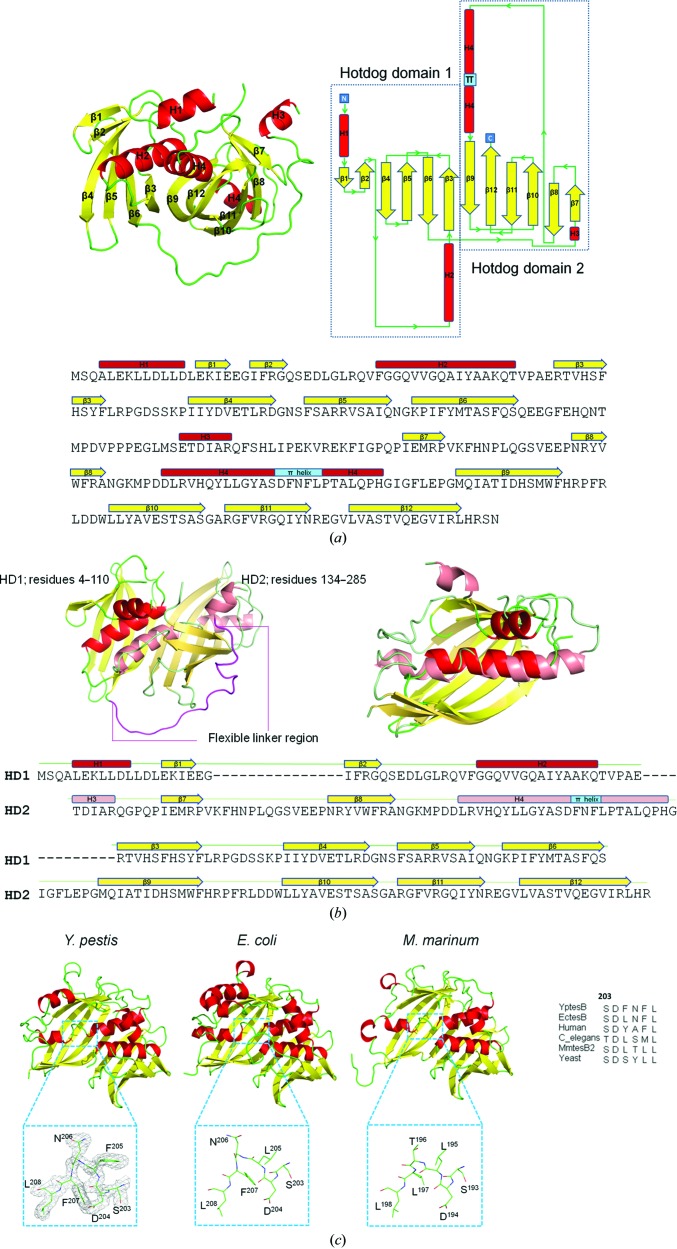
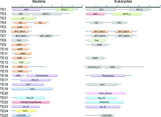
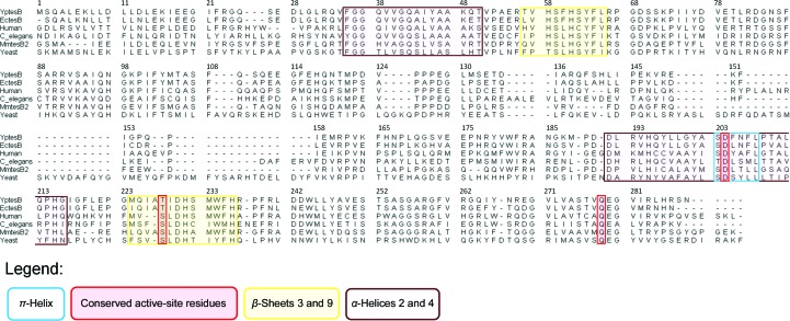
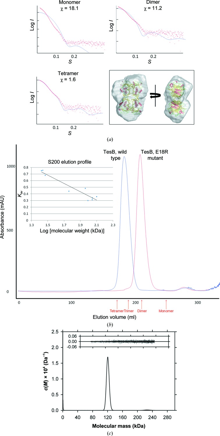
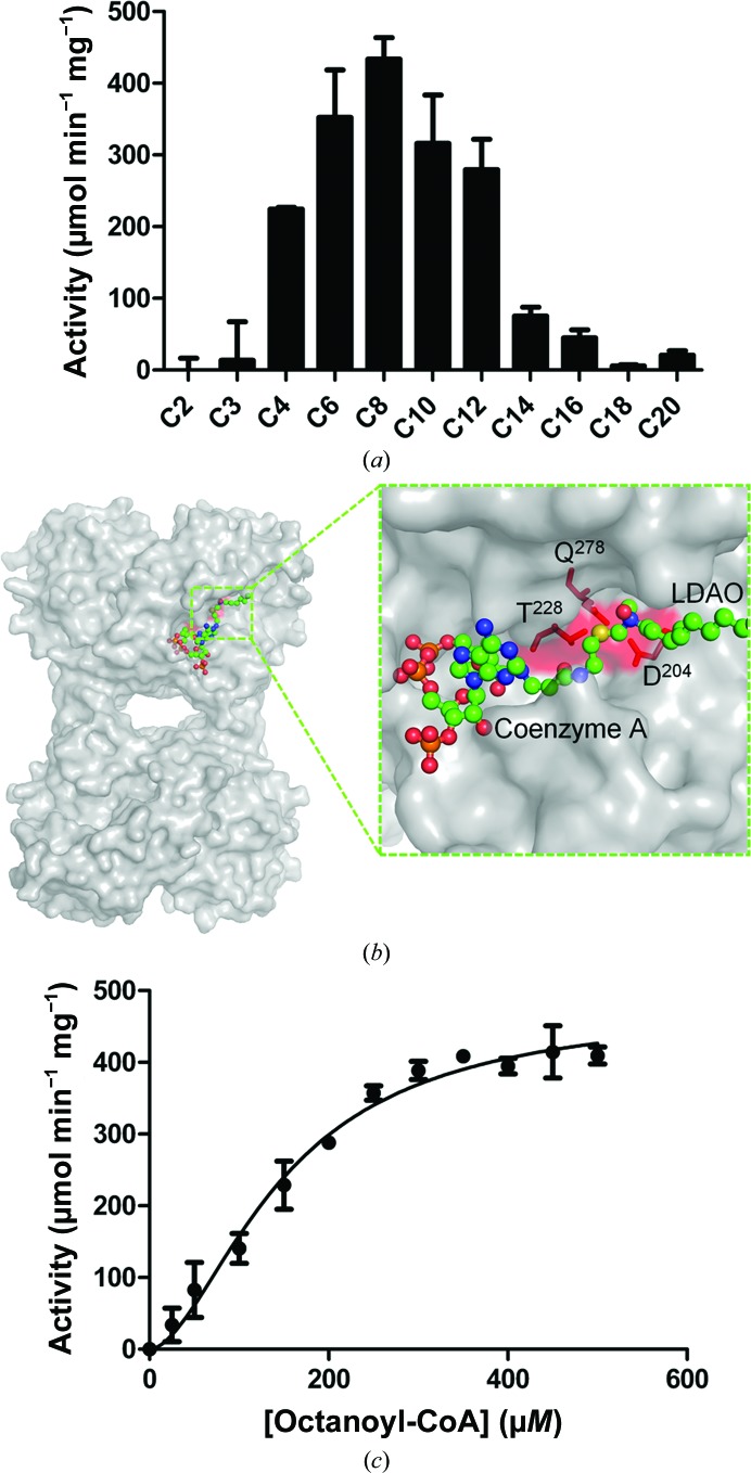
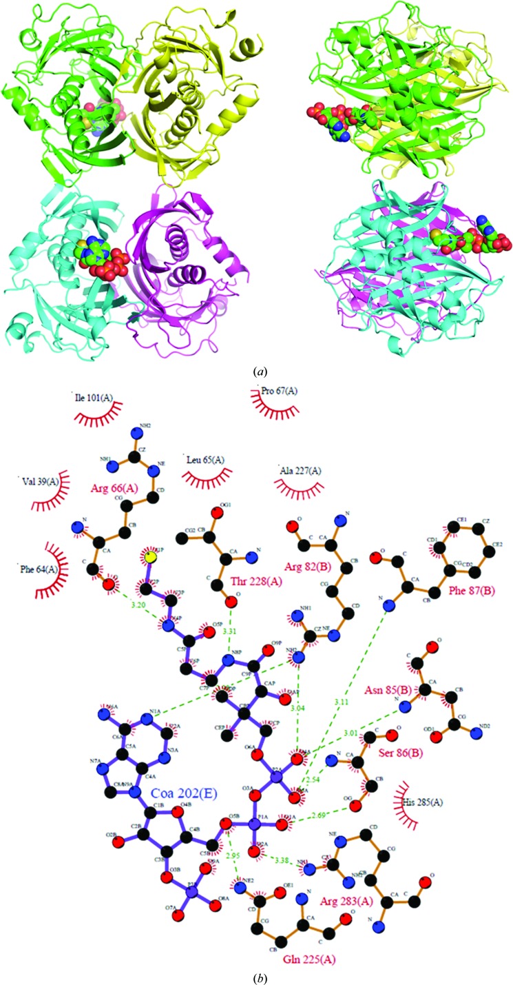
Acyl-CoA thioesterases catalyse the hydrolysis of the thioester bonds present within a wide range of acyl-CoA substrates, releasing free CoASH and the corresponding fatty-acyl conjugate. The TesB-type thioesterases are members of the TE4 thioesterase family, one of 25 thioesterase enzyme families characterized to date, and contain two fused hotdog domains in both prokaryote and eukaryote homologues. Only two structures have been elucidated within this enzyme family, and much of the current understanding of the TesB thioesterases has been based on the Escherichia coli structure. Yersinia pestis, a highly virulent bacterium, encodes only one TesB-type thioesterase in its genome; here, the structural and functional characterization of this enzyme are reported, revealing unique elements both within the protomer and quaternary arrangements of the hotdog domains which have not been reported previously in any thioesterase family. The quaternary structure, confirmed using a range of structural and biophysical techniques including crystallography, small-angle X-ray scattering, analytical ultracentrifugation and size-exclusion chromatography, exhibits a unique octameric arrangement of hotdog domains. Interestingly, the same biological unit appears to be present in both TesB structures solved to date, and is likely to be a conserved and distinguishing feature of TesB-type thioesterases. Analysis of the Y. pestis TesB thioesterase activity revealed a strong preference for octanoyl-CoA and this is supported by structural analysis of the active site. Overall, the results provide novel insights into the structure of TesB thioesterases which are likely to be conserved and distinguishing features of the TE4 thioesterase family.

Keywords: TesB; Yersinia pestis; acyl-CoA thioesterases.


Figures

Figure 1
Figure 1
The primary, secondary and tertiary structure of TesB with α-helices coloured red, β-strands yellow and loops green. (a) The double hotdog-domain structure of YpTesB. (b) The two hotdog domains of YpTesB, with a superposition of the domains (r.m.s.d. of 0.33 Å) and sequence alignment, and (c) a comparison of three TesB structures from Y. pestis (PDB entry 4qfw), E. coli (PDB entry 1c8u) and M. marinum (PDB entry 3u0a), revealing a conserved β-bulge through the HD2 α-helix.
Figure 2
Figure 2
Domain architecture of thioesterase families as presented in the ThYme database (http://www.enzyme.cbirc.iastate.edu/). Abbreviations: ACH, acetyl-CoA hydrolase; ACH_C, acetyl-CoA hydrolase C superfamily; BHT, bile hydrolase transferase; LL1, lysophospholipase L1-like; TEII, thioesterase II; PaaI, phenylacetic acid thioesterase; PPB, phosphopantetheine-binding domain; PKS_TE, polyketide synthase thioesterase; GrsT, gramicidin S biosynthesis thioesterase; luxD, lux-specific myristoyl-ACP thioesterase; Pep_S9, peptidase_S9 superfamily; Lac_B, lactamase_B superfamily. Domains presented in grey have no solved structures and are therefore theoretical.
Figure 3
Figure 3
The primary sequence of the π-helix (in blue) is conserved throughout these proteins and spans Asp204 of the active site.
Figure 4
Figure 4
(a) The quaternary structure of TesB is a tetramer of protomers with a Glu residue mutated to disrupt the tetramer configuration (inset) which is conserved within the E. coli and M. marinum structures (b).
Figure 5
Figure 5
The tetramer was confirmed to be the biological unit using a number of biophysical techniques. (a) Small-angle X-ray scattering data were compared with scattering data generated using CRYSOL (Svergun et al., 1995 ▶) for a monomer, a dimer and a tetramer, with the best fit for the tetramer, and a SAXS envelope was generated using the experimental data. (b) YpTesB eluted from a size-exclusion column consistent with a tetramer, as confirmed using a standard curve of the size-exclusion column (inset) to determine the elution volumes of a monomer, a dimer, a trimer and a tetramer (red). (c) The continuous mass [c(M)] distribution is plotted as a function of molecular mass (kDa) for TesB (2.4 mg ml−1). The molecular mass at the ordinate maximum of the peak shown corresponds to 120 kDa. The c(M) distribution was calculated using 200 masses from 0 to 300 kDa at a P-value of 0.95, which resulted in an r.m.s.d. of 0.00685 and a runs test Z of 7.61 and yielded a frictional ratio of 1.28. Inset: residuals for the c(M) best fit plotted as a function of radial position.
Figure 6
Figure 6
TesB activity against a range of substrates (a), demonstrating activity against a broad range of fatty acyl-CoA chain lengths; octanoyl-CoA was identified as the preferred substrate and was investigated further. (b) The active site with CoA and LDAO (from the EcTesB model) superimposed. (c) Activity curve for octanoyl-CoA, with a Hill coefficient of 1.753 and a V max of 478 µmol min−1 mg−1.
Figure 7
Figure 7
(a) The structure of TesB solved in the presence of CoA; the same octameric configuration is observed as for the apo form of the enzyme. (b) LigPlot representation of the detailed interactions of CoA with TesB (Wallace et al., 1995 ▶).

References

    1. Cabanel, N., Leclercq, A., Chenal-Francisque, V., Annajar, B., Rajerison, M., Bekkhoucha, S., Bertherat, E. & Carniel, E. (2013). Emerg. Infect. Dis. 19, 230–236. - PMC - PubMed
    1. Cantu, D. C., Chen, Y. & Reilly, P. J. (2010). Protein Sci. 19, 1281–1295. - PMC - PubMed
    1. Cooley, R. B., Arp, D. J. & Karplus, P. A. (2010). J. Mol. Biol. 404, 232–246. - PMC - PubMed
    1. Dias, M. V., Huang, F., Chirgadze, D. Y., Tosin, M., Spiteller, D., Dry, E. F., Leadlay, P. F., Spencer, J. B. & Blundell, T. L. (2010). J. Biol. Chem. 285, 22495–22504. - PMC - PubMed
    1. Dillon, S. C. & Bateman, A. (2004). BMC Bioinformatics, 5, 109. - PMC - PubMed

Publication types