. 2015 Apr 2;16(4):7377-93.
doi: 10.3390/ijms16047377.

Role of glucokinase in the subcellular localization of glucokinase regulatory protein

Affiliations

Role of glucokinase in the subcellular localization of glucokinase regulatory protein

Ling Jin et al. Int J Mol Sci. .

Abstract

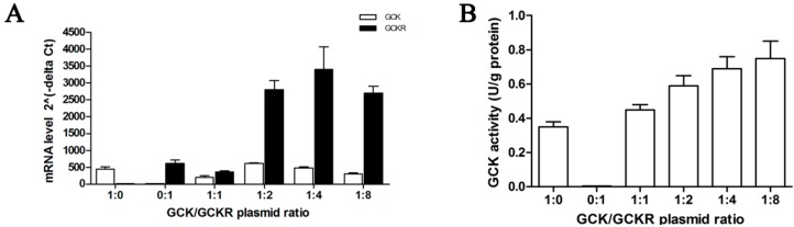
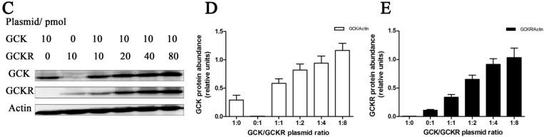
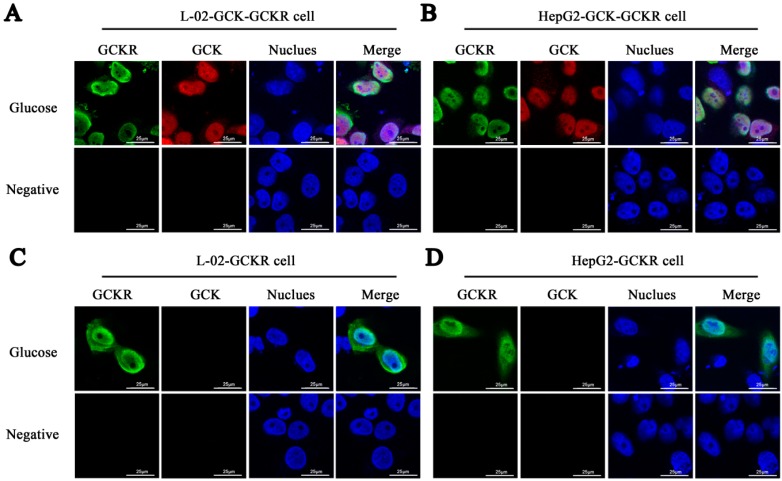
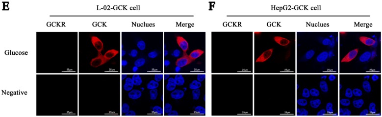
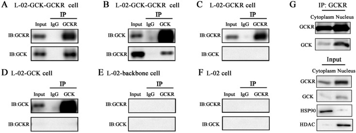
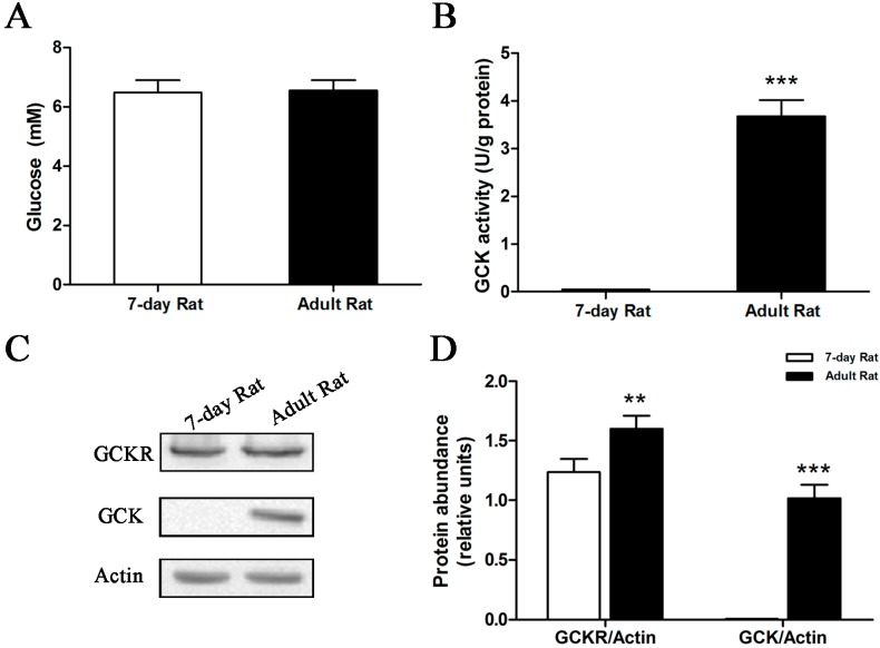
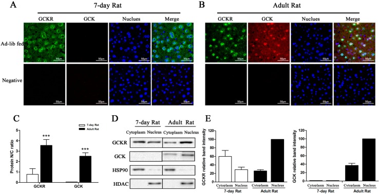
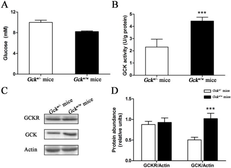
Glucokinase (GCK) is the rate-limiting enzyme of liver glucose metabolism. Through protein-protein interactions, glucokinase regulatory protein (GCKR) post-transcriptionally regulates GCK function in the liver, and causes its nuclear localization. However the role of GCK in regulating GCKR localization is unknown. In the present study, using in vitro and in vivo models, we examined the levels of GCK and GCKR, and their subcellular localization. We found that total cellular levels of GCKR did not vary in the in vivo models, but its subcellular localization did. In animals with normal levels of GCK, GCKR is mainly localized to the nuclei of hepatocytes. In seven-day old rats and liver-specific Gck gene knockout mice (animals that lack or have reduced levels of GCK protein), GCKR was found primarily in the cytoplasm. The interaction of GCK with GCKR was further examined using in vitro models where we varied the levels of GCK and GCKR. Varying the level of GCK protein had no effect on total cellular GCKR protein levels. Taken together, our results indicate that GCK is important for the localization of GCKR to the nucleus and raises the possibility that GCKR may have functions in addition to those regulating GCK activity in the cytoplasm.


Figures

Figure 1
Figure 1
Expression of glucokinase (GCK) and glucokinase regulatory protein (GCKR) in co-transfected liver L-02 cells. GCK and GCKR expressing plasmids were co-transfected into L-02 cells at a range of plasmid ratios (molar) from 1:1 to 1:8. Cells transfected with the GCK expressing, GCKR expressing, and GCKR backbone plasmids were used as controls. (A) Quantitative real-time RT-PCR assessment of the mRNA levels of Gck (open bars) and Gckr (solid bars) at different plasmid ratios; (B) GCK enzymatic activity at different plasmid ratios; (C) Representative Western blots for GCK and GCKR, with β-actin used as a loading control, at different plasmid ratios; and (D,E) Quantification of the immunoblots for GCK (D) and GCKR (E), with relative units for GCK (D) or GCKR (E) abundance being the abundance of GCK or GCKR normalized to the β-actin level for that sample. Data are presented as means ± S.D. (Standard Deviation) (n = 4).
Figure 1
Figure 1
Expression of glucokinase (GCK) and glucokinase regulatory protein (GCKR) in co-transfected liver L-02 cells. GCK and GCKR expressing plasmids were co-transfected into L-02 cells at a range of plasmid ratios (molar) from 1:1 to 1:8. Cells transfected with the GCK expressing, GCKR expressing, and GCKR backbone plasmids were used as controls. (A) Quantitative real-time RT-PCR assessment of the mRNA levels of Gck (open bars) and Gckr (solid bars) at different plasmid ratios; (B) GCK enzymatic activity at different plasmid ratios; (C) Representative Western blots for GCK and GCKR, with β-actin used as a loading control, at different plasmid ratios; and (D,E) Quantification of the immunoblots for GCK (D) and GCKR (E), with relative units for GCK (D) or GCKR (E) abundance being the abundance of GCK or GCKR normalized to the β-actin level for that sample. Data are presented as means ± S.D. (Standard Deviation) (n = 4).
Figure 2
Figure 2
Sub-cellular localization of GCK and GCKR in liver L-02 and HepG2 cells in glucose containing media. GCK and/or GCKR expressing plasmids were transfected into liver cell lines. Forty-eight hours after transfection, culture medium was changed to low glucose (5.5 mM glucose). Confocal immunofluoresence micrographs are shown for cells after culturing in low glucose for 2 h. (A) L-02 cells co-transfected with GCK and GCKR plasmids; (B) HepG2 cells co-transfected with GCK and GCKR plasmids; (C) L-02 cells transfected with GCKR plasmids; (D) HepG2 cells transfected with GCKR plasmids; (E) L-02 cells transfected with GCK plasmids; and (F) HepG2 cells transfected with GCK plasmids. GCKR (revealed in green) and GCK (revealed in red) were detected using antibodies, and the nuclei were counterstained with Hoechst (stained in blue). The overlap of the three channels is shown as Merge. White color in the merged panels in (A) and (B) denotes co-localization of GCKR and GCK in the nucleus. Cells incubated with only secondary antibodies were used as negative controls. Images are representative of four experiments with similar results. Scale bar = 25 μm.
Figure 2
Figure 2
Sub-cellular localization of GCK and GCKR in liver L-02 and HepG2 cells in glucose containing media. GCK and/or GCKR expressing plasmids were transfected into liver cell lines. Forty-eight hours after transfection, culture medium was changed to low glucose (5.5 mM glucose). Confocal immunofluoresence micrographs are shown for cells after culturing in low glucose for 2 h. (A) L-02 cells co-transfected with GCK and GCKR plasmids; (B) HepG2 cells co-transfected with GCK and GCKR plasmids; (C) L-02 cells transfected with GCKR plasmids; (D) HepG2 cells transfected with GCKR plasmids; (E) L-02 cells transfected with GCK plasmids; and (F) HepG2 cells transfected with GCK plasmids. GCKR (revealed in green) and GCK (revealed in red) were detected using antibodies, and the nuclei were counterstained with Hoechst (stained in blue). The overlap of the three channels is shown as Merge. White color in the merged panels in (A) and (B) denotes co-localization of GCKR and GCK in the nucleus. Cells incubated with only secondary antibodies were used as negative controls. Images are representative of four experiments with similar results. Scale bar = 25 μm.
Figure 3
Figure 3
Interaction of GCK and GCKR in whole cell lysates, and nuclear fractions, of co-transfected L-02 cells. L-02 cells were transfected with GCKR and/or GCK expressing plasmids. Total protein cellular lysates were immunoprecipitated with anti-GCKR (A,C,E,F) or anti-GCK (B,D) followed by immunoblotting with anti-GCK and anti-GCKR. Cytoplasmic and nuclear protein cellular lysates were immunoprecipitated with anti-GCKR followed by immunoblotting with anti-GCK and anti-GCKR (A,G). Immunoprecipitation of GCKR in the co-transfected L-02 cells; (B) Immunoprecipitaion of GCK in the co-transfected L-02 cells; (C) Immunoprecipitation of GCKR in GCKR plasmid transfected cells; (D) Immunoprecipitation of GCK in GCK plasmid transfected cells; (E,F) No immunoprecipitation of GCK and GCKR in cells transfected by the backbone vector (E) and in untransfected L-02 cells (F,G). Cytoplasmic and nuclear immunoprecipitation of GCKR in the co-transfected L-02 cells. Hsp90 and histone deacetylase (HDAC) were used as cytoplasmic and nuclear fraction markers, respectively. Input represents cell lysates before co-IP. Data are representative of four experiments with similar results.
Figure 4
Figure 4
Glucokinase activity and protein levels in seven-day old rats. Rats, seven-day old and adults, had free access to food and water. (A) Serum glucose concentrations; (B) GCK enzymatic activity from liver extracts; (C) Representative Western blot of GCK and GCKR protein levels, with β-actin used as a loading control; and (D) Quantification of the GCK and GCKR immunoblots, for seven-day old and adult rats. Relative units for GCK and GCKR protein abundance in (D) are the abundance of GCK or GCKR normalized to the β-actin levels of that sample. ** p < 0.01, *** p < 0.001 vs. seven-day old rat. Data are presented as means ± S.D. (n = 4).
Figure 5
Figure 5
Sub-cellular localization of GCKR in seven-day old rats. (A,B) Confocal immunofluoresence micrographs with antibodies against GCKR (revealed in green), GCK (revealed in red) with nuclei counterstained with Hoechst (stained in blue) in seven-day old (A) and adult (B) rats. Overlap of the three channels is shown in Merge. White color in the merged panels denotes co-localization of GCKR and GCK in the nucleus. Slides incubated with only the secondary antibodies were used as negative controls. Images are representative of four experiments with similar results. Scale bar = 50 μm; (C) Nuclear/cytoplasmic ratio (N/C ratio) for GCKR and GCK derived from immunoflouresence. Data are presented as means ± S.D. from four individual hepatocyte preparations with 30 cells in total. *** p < 0.001 vs. 7-day old rat. (D) Representative Western blots for GCKR and GCK protein from the cytosolic (C) and nuclear (N) fractions. Abundance of GCK and GCKR in the cytosolic and nuclear fractions was normalized for comparable amounts of protein. Hsp90 and HDAC are markers for the cytoplasmic and nuclear fractions, respectively; (E) Densitometry analysis of the immunoblots in (D). Intensity of the protein band in the nuclear fraction of the adult rat is set as 100. Data are representative of four experiments with similar results.
Figure 6
Figure 6
Enzymatic and protein levels for glucokinase in the liver-specific Gck gene knockout (Gckw/−) mice. Ad libitum fed mice had free access to food and water. (A) Serum glucose concentrations; (B) GCK activity in the liver; (C) Representative Western blots for GCK and GCKR, with β-actin as a loading control; and (D) quantification of the immunoblots of glucokinase and GCKR are shown for Gckw/− and Gckw/w mice in the ad libitum fed state Relative units for GCK and GCKR protein abundance in (D) are the abundance of GCK or GCKR normalized to the β-actin level of that sample. *** p < 0.001 vs. Gckw/− mice. Data are presented as means ± S.D. (n = 4).
Figure 7
Figure 7
Sub-cellular localization of GCKR in the liver-specific Gck gene knockout (Gckw/−) mice. (A,B) Confocal immunofluoresence micrographs with antibodies against GCKR (revealed in green), GCK (revealed in red) with the nuclei counterstained with Hoechst (stained in blue) in (Gckw/−) (A) and wild-type (Gckw/w) (B) mice. Overlap of the three channels is shown in Merge. White color in the merged panels denotes co-localization of GCKR and GCK in the nucleus. Slides incubated with only the secondary antibodies were used as negative controls. Images are representative of four experiments with similar results. Scale bar = 50 μm; (C) Nuclear/cytoplasmic ratios (N/C ratio) for GCKR and GCK are based on the immunoflouresence. Data are presented as means ± S.D. from four individual hepatocyte preparations with 30 cells in total. * p < 0.05, *** p < 0.001 vs. Gckw/− mice; (D) Representative Western blots for GCKR and GCK protein in the cytosolic (C) and nuclear (N) fractions. Cytosolic and nuclear fractions are normalized for comparable amounts of protein. Hsp90 and HDAC were used as cytoplasmic and nuclear fraction markers, respectively; (E) Densitometry analysis of the immunoblot is shown in (D). Relative band intensity of the proteins in the nuclear fraction of the ad libitum fed wild-type mice is set as 100. Data are representative of four experiments with similar results.

Similar articles

Cited by

References

    1. Vandercammen A., van Schaftingen E. Species and tissue distribution of the regulatory protein of glucokinase. Biochem. J. 1993;294:551–556. - PMC - PubMed
    1. Van Schaftingen E. A protein from rat liver confers to glucokinase the property of being antagonistically regulated by fructose 6-phosphate and fructose 1-phosphate. Eur. J. Biochem. 1989;179:179–184. - PubMed
    1. Vandercammen A., van Schaftingen E. The mechanism by which rat liver glucokinase is inhibited by the regulatory protein. Eur. J. Biochem. 1990;191:483–489. doi: 10.1111/j.1432-1033.1990.tb19147.x. - DOI - PubMed
    1. Farrelly D., Brown K.S., Tieman A., Ren J., Lira S.A., Hagan D., Gregg R., Mookhtiar K.A., Hariharan N. Mice mutant for glucokinase regulatory protein exhibit decreased liver glucokinase: A sequestration mechanism in metabolic regulation. Proc. Natl. Acad. Sci. USA. 1999;96:14511–14516. doi: 10.1073/pnas.96.25.14511. - DOI - PMC - PubMed
    1. Grimsby J., Coffey J.W., Dvorozniak M.T., Magram J., Li G., Matschinsky F.M., Shiota C., Kaur S., Magnuson M.A., Grippo J.F. Characterization of glucokinase regulatory protein-deficient mice. J. Boil. Chem. 2000;275:7826–7831. doi: 10.1074/jbc.275.11.7826. - DOI - PubMed

Publication types

LinkOut - more resources