. 2015 Apr 7;10(4):e0122634.
doi: 10.1371/journal.pone.0122634. eCollection 2015.

Mitogen-activated protein kinases are associated with the regulation of physiological traits and virulence in Fusarium oxysporum f. sp. cubense

Affiliations

Mitogen-activated protein kinases are associated with the regulation of physiological traits and virulence in Fusarium oxysporum f. sp. cubense

Zhaojian Ding et al. PLoS One. .

Abstract

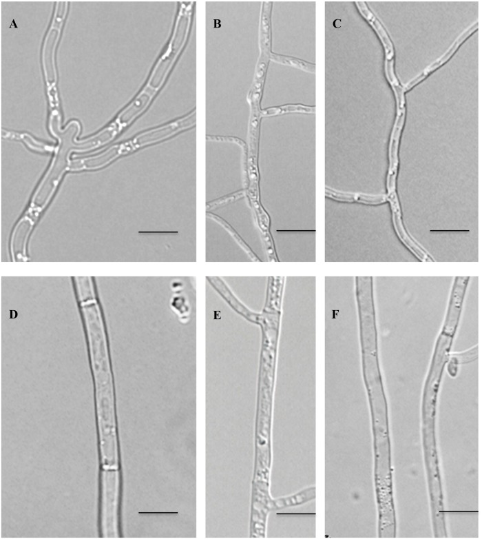
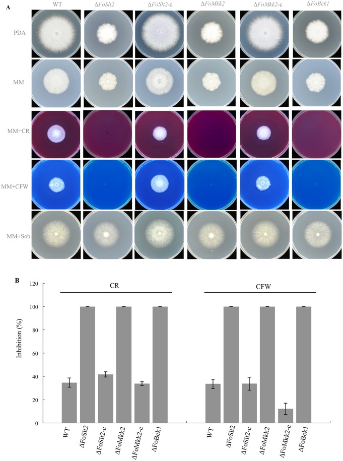
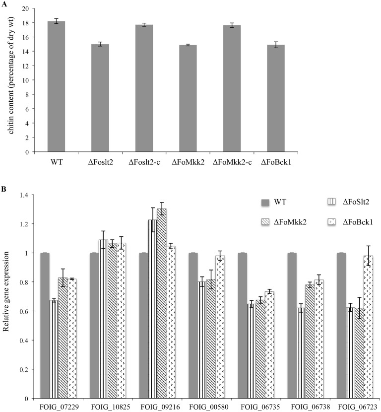
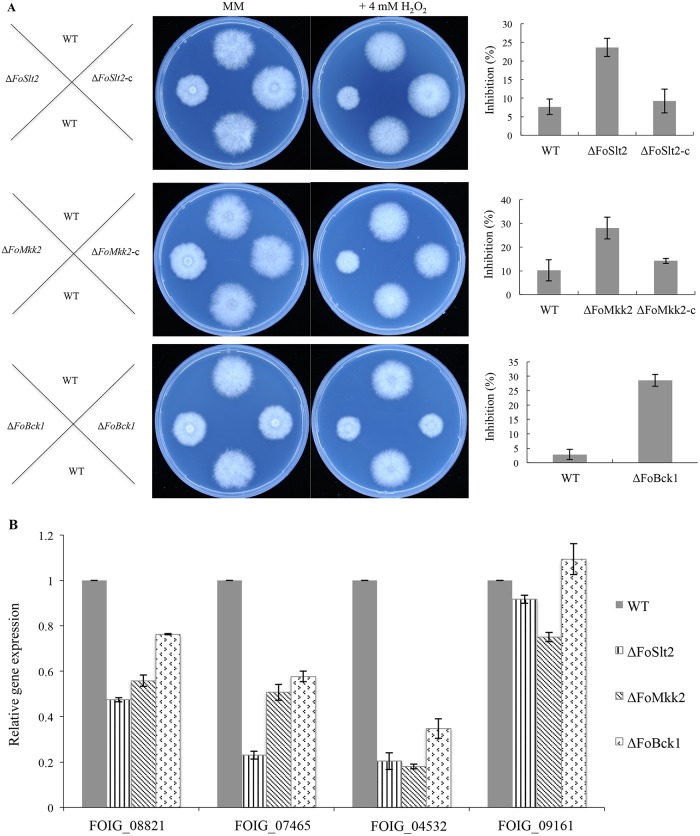
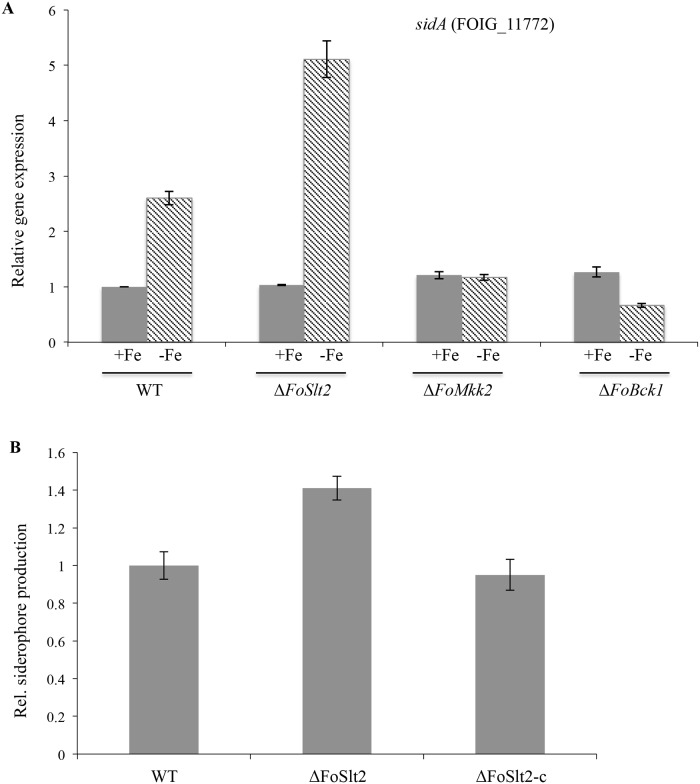
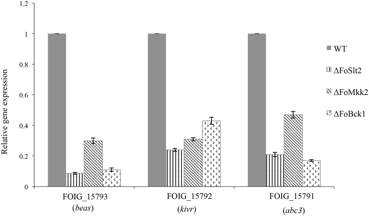
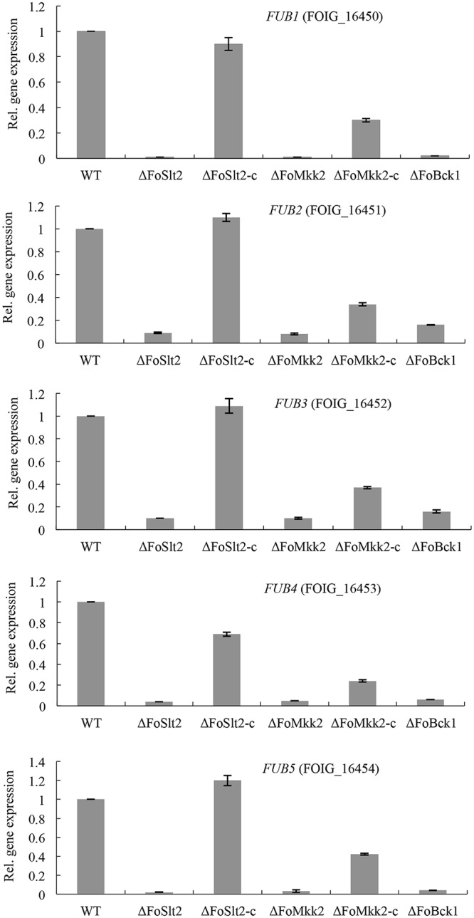
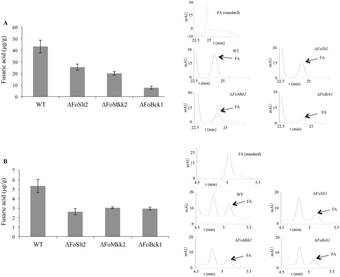
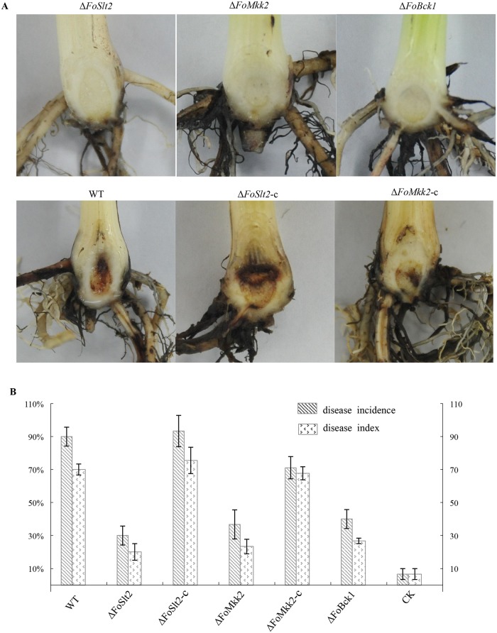
Fusarium oxysporum f. sp. cubense (FOC) is an important soil-borne fungal pathogen causing devastating vascular wilt disease of banana plants and has become a great concern threatening banana production worldwide. However, little information is known about the molecular mechanisms that govern the expression of virulence determinants of this important fungal pathogen. In this study, we showed that null mutation of three mitogen-activated protein (MAP) kinase genes, designated as FoSlt2, FoMkk2 and FoBck1, respectively, led to substantial attenuation in fungal virulence on banana plants. Transcriptional analysis revealed that the MAP kinase signaling pathway plays a key role in regulation of the genes encoding production of chitin, peroxidase, beauvericin and fusaric acid. Biochemical analysis further confirmed the essential role of MAP kinases in modulating the production of fusaric acid, which was a crucial phytotoxin in accelerating development of Fusarium wilt symptoms in banana plants. Additionally, we found that the MAP kinase FoSlt2 was required for siderophore biosynthesis under iron-depletion conditions. Moreover, disruption of the MAP kinase genes resulted in abnormal hypha and increased sensitivity to Congo Red, Calcofluor White and H2O2. Taken together, these results depict the critical roles of MAP kinases in regulation of FOC physiology and virulence.


Conflict of interest statement

Competing Interests: The authors have declared that no competing interests exist.

Figures

Fig 1
Fig 1. Hypha morphology of WT, mutants ΔFoSlt2, ΔFoMkk2 and ΔFoBck1, and complemented strains ΔFoSlt2-c and ΔFoMkk2-c.
Strains were incubated at 28°C for 4 days on PDA plates. A: ΔFoSlt2, B: ΔFoMkk2, C: ΔFoBck1, D: WT, E: ΔFoSlt2-c, F: ΔFoMkk2-c. Bars: 10μm.
Fig 2
Fig 2. Effects of FoSlt2, FoMkk2 and FoBck1 genes on cell wall integrity of FOC.
(A) Colony morphology of the indicated strains grown on PDA, minimal medium (MM), MM supplemented with Congo Red (CR, 40 μg/mL), Calcofluor White (CFW, 40 μg/mL), or sorbitol (Sob, 1.2 M) incubated at 28°C for 6 days on MM plates. (B) Inhibition of the radiated growth of the indicated strains grown on the MM plates. Error bars indicate the standard error from three replicates.
Fig 3
Fig 3. Quantification of chitin and expression of chitin synthase genes.
(A) Chitin content in WT, mutants ΔFoSlt2, ΔFoMkk2 and ΔFoBck1, complemented strains ΔFoSlt2-c and ΔFoMkk2-c. (B) Quantitative real-time PCR analysis of chitin synthase in WT, mutants ΔFoSlt2, ΔFoMkk2 and ΔFoBck1. The seven chitin synthase are FOIG_07229, FOIG_10825, FOIG_09216, FOIG_00580, FOIG_06735, FOIG_06738 and FOIG_06723. Error bars indicate the standard error from three replicates.
Fig 4
Fig 4. The deletion mutants were sensitive to H2O2.
(A) Middle panels of figures show colony morphology of the WT, mutants ΔFoSlt2, ΔFoMkk2 and ΔFoBck1, complemented strains ΔFoSlt2-c and ΔFoMkk2-c were incubated on MM plates supplemented with 4 mM H2O2 at 28°C for 3.5 days. Right panels of figures show inhibition of the radiated growth of the indicated strains grown on the MM plates with the same order as the middle figures. (B) Quantitative real-time PCR analysis of peroxidase synthase genes in the WT, mutants ΔFoSlt2, ΔFoMkk2 and ΔFoBck1. The four peroxidase synthase genes are FOIG_08821, FOIG_07465, FOIG_04532 and FOIG_09161. Error bars indicate the standard error from three replicates.
Fig 5
Fig 5. Determination of siderophore biosynthesis in mutant ΔFoSlt2 during iron-poor conditions.
(A) The siderophore biosynthetic gene sidA was upregulated in mutant ΔFoSlt2 during iron-poor conditions, normalized to WT using quantitative real-time PCR. (B) CAS assay showed increased level of secreted siderophores in mutant ΔFoSlt2 during iron-poor conditions, normalized to WT. The indicated strains were incubated for 5 days and the supernatants were analyzed for siderophore content. Error bars indicate the standard error from three replicates.
Fig 6
Fig 6. The MAP kinases affect the transcription of beauvericin biosynthetic genes.
Gene expression levels are represented relative to WT using quantitative real-time PCR. Error bars indicate the standard error from three replicates.
Fig 7
Fig 7. The MAP kinases regulate transcription of the fusaric acid gene cluster.
Gene expression levels are represented relative to WT using quantitative real-time PCR. Error bars indicate the standard error from three replicates.
Fig 8
Fig 8. Fusaric acid (FA) production was reduced in mutants ΔFoSlt2, ΔFoMkk2 and ΔFoBck1.
(A) Fusaric acid production from cultures of the indicated strains in Czapek dox medium was analysed by High Performance Liquid Chromatography (HPLC). Fusaric acid levels are represented in μg g-1 mycelial dry weight. (B) Fusaric acid production from the crude mycotoxin extracted of the indicated strains in grains cultures was analysed with HPLC. Fusaric acid levels are represented in μg g-1 grains cultures dry weight. Error bars indicate the standard error from three replicates.
Fig 9
Fig 9. Virulence assay of WT, mutants ΔFoSlt2, ΔFoMkk2 and ΔFoBck1, and complemented strains ΔFoSlt2-c and ΔFoMkk2-c.
(A) Disease symptom on corms of banana tissue plants were assayed after 40 days of inoculation with indicated strains and water as negative control. (B) Disease incidence and disease index indicated disease severity. Error bars indicate the standard error from three replicates.

Similar articles

Cited by

References

    1. Ploetz RC (2006) Fusarium wilt of banana is caused by several pathogens referred to as Fusarium oxysporum f. sp cubense . Phytopathology 96: 653–656. 10.1094/PHYTO-96-0653 - DOI - PubMed
    1. Li MH, Xie XL, Lin XF, Shi JX, Ding ZJ, Ling JF, et al. (2014) Functional characterization of the gene FoOCH1 encoding a putative α-1,6-mannosyltransferase in Fusarium oxysporum f. sp. cubense . Fungal Genet Biol 65: 1–13. 10.1016/j.fgb.2014.01.005 - DOI - PubMed
    1. Qi XZ, Guo LJ, Yang LY, Huang JS (2013) Foatf1, a bZIP transcription factor of Fusarium oxysporum f. sp cubense, is involved in pathogenesis by regulating the oxidative stress responses of Cavendish banana (Musa spp.). Physiol Mol Plant P 84: 76–85.
    1. Zhao X, Kim Y, Park G, Xu JR (2005) A mitogen-activated protein kinase cascade regulating infection-related morphogenesis in Magnaporthe grisea . Plant Cell 17: 1317–1329. - PMC - PubMed
    1. Rispail N, Soanes DM, Ant C, Czajkowski R, Grunler A, Huguet R, et al. (2009) Comparative genomics of MAP kinase and calcium-calcineurin signalling components in plant and human pathogenic fungi. Fungal Genet Biol 46: 287–298. 10.1016/j.fgb.2009.01.002 - DOI - PubMed

Publication types

LinkOut - more resources