. 2015 Apr 7;10(4):e0123293.
doi: 10.1371/journal.pone.0123293. eCollection 2015.

Oxidation by neutrophils-derived HOCl increases immunogenicity of proteins by converting them into ligands of several endocytic receptors involved in antigen uptake by dendritic cells and macrophages

Affiliations

Oxidation by neutrophils-derived HOCl increases immunogenicity of proteins by converting them into ligands of several endocytic receptors involved in antigen uptake by dendritic cells and macrophages

Rafał Biedroń et al. PLoS One. .

Erratum in

Abstract

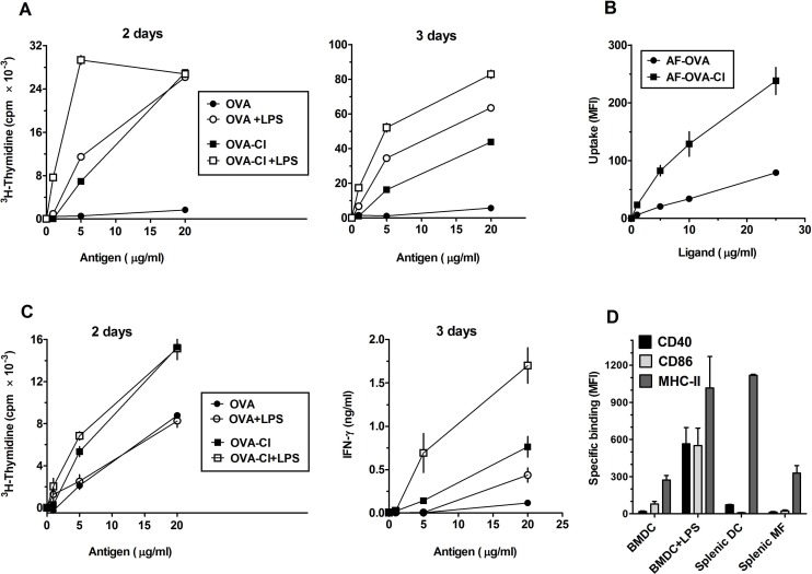
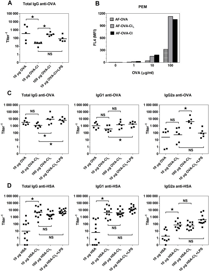
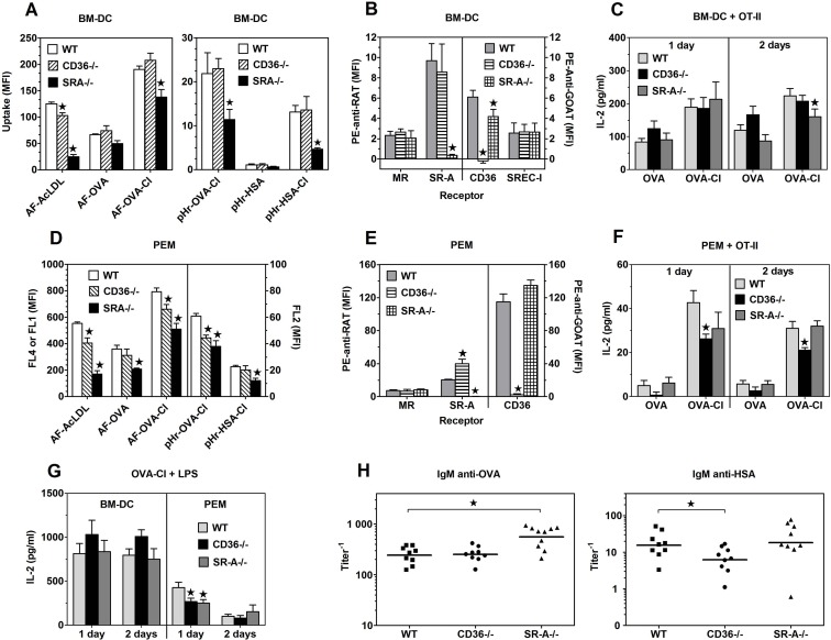
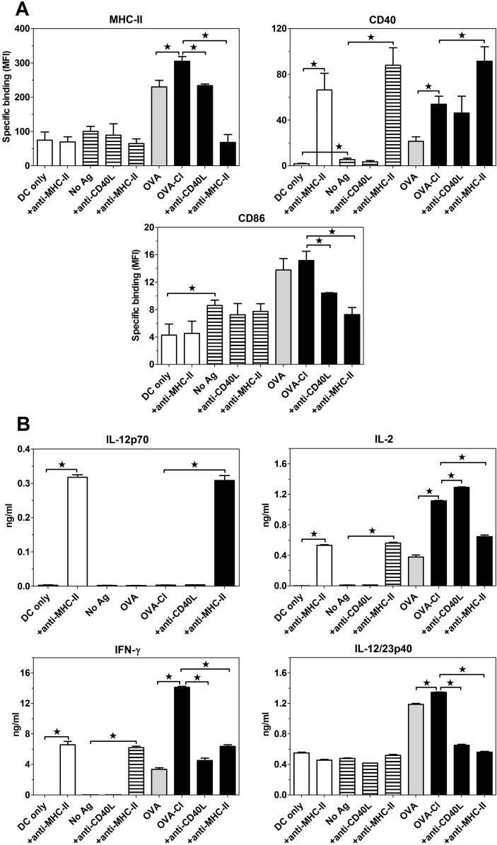
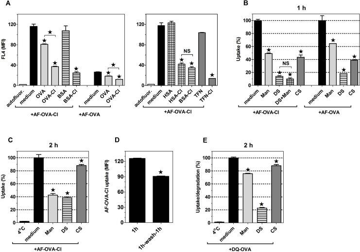
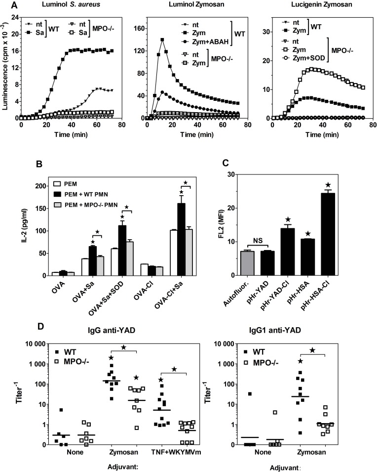
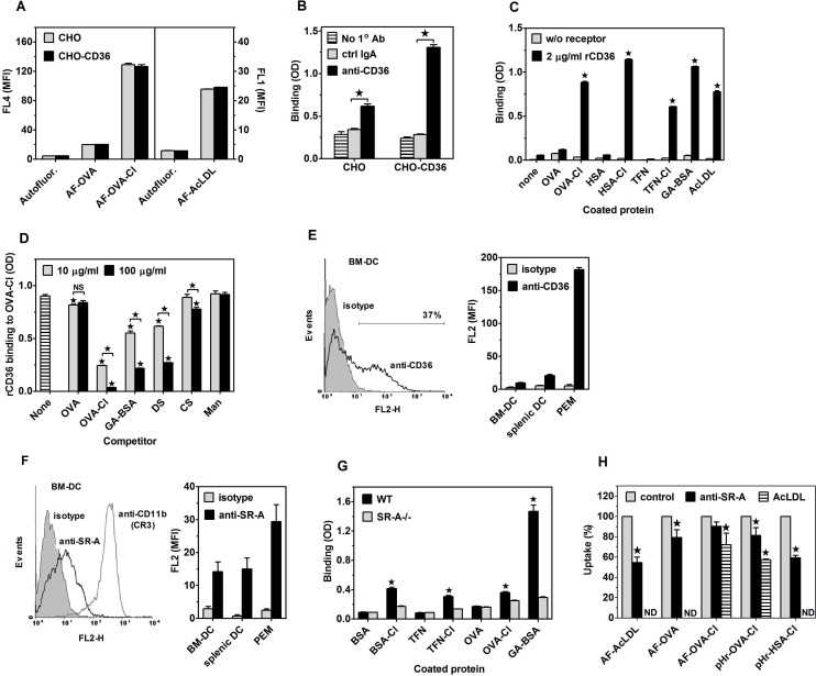
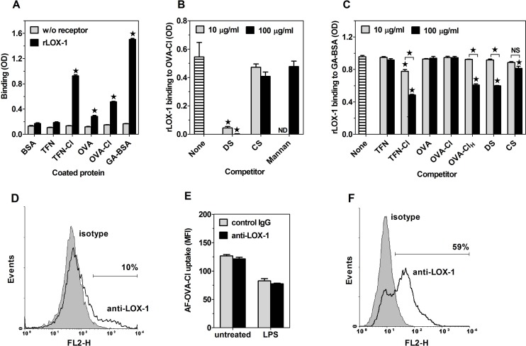
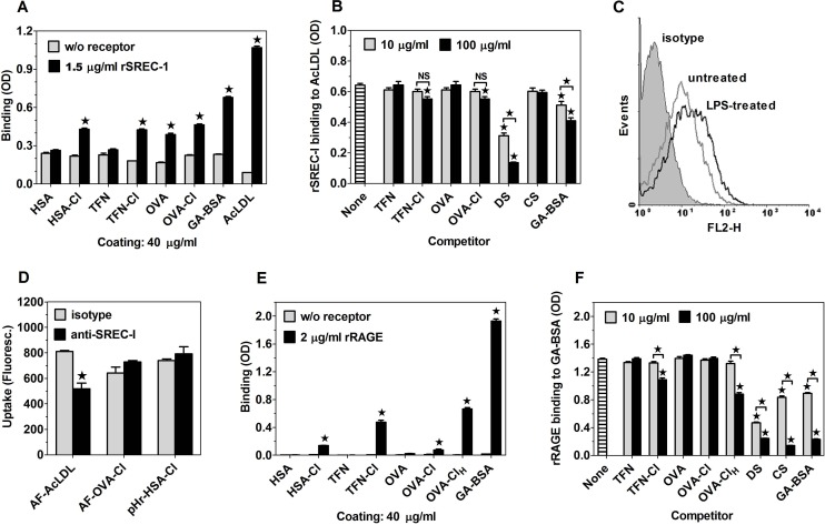
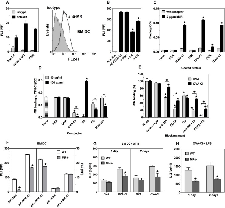
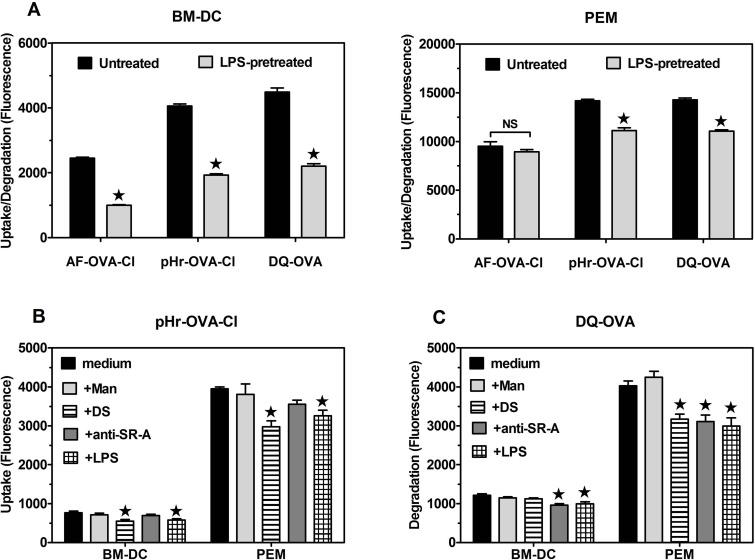
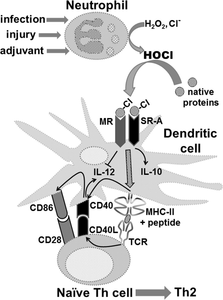
The initiation of adaptive immune responses to protein antigens has to be preceded by their uptake by antigen presenting cells and intracellular proteolytic processing. Paradoxically, endocytic receptors involved in antigen uptake do not bind the majority of proteins, which may be the main reason why purified proteins stimulate at most weak immune responses. A shared feature of different types of adjuvants, capable of boosting immunogenicity of protein vaccines, is their ability to induce acute inflammation, characterized by early influx of activated neutrophils. Neutrophils are also rapidly recruited to sites of tissue injury or infection. These cells are the source of potent oxidants, including hypochlorous acid (HOCl), causing oxidation of proteins present in inflammatory foci. We demonstrate that oxidation of proteins by endogenous, neutrophils-derived HOCl increases their immunogenicity. Upon oxidation, different, randomly chosen simple proteins (yeast alcohol dehydrogenase, human and bovine serum albumin) and glycoproteins (human apo-transferrin, ovalbumin) gain the ability to bind with high affinity to several endocytic receptors on antigen presenting cells, which seems to be the major mechanism of their increased immunogenicity. The mannose receptor (CD206), scavenger receptors A (CD204) and CD36 were responsible for the uptake and presentation of HOCl-modified proteins by murine dendritic cells and macrophages. Other scavenger receptors, SREC-I and LOX-1, as well as RAGE were also able to bind HOCl-modified proteins, but they did not contribute significantly to these ligands uptake by dendritic cells because they were either not expressed or exhibited preference for more heavily oxidised proteins. Our results indicate that oxidation by neutrophils-derived HOCl may be a physiological mechanism of conferring immunogenicity on proteins which in their native forms do not bind to endocytic receptors. This mechanism might enable the immune system to detect infections caused by pathogens not recognized by pattern recognition receptors.


Conflict of interest statement

Competing Interests: The authors have declared that no competing interests exist.

Figures

Fig 1
Fig 1. OVA-Cl exhibits increased immunogenicity that may be caused by enhanced uptake by APC.
(A) BM-DC were incubated with indicated concentrations of OVA or OVA-Cl for 2 h at 37°C. When indicated, 200 ng/ml LPS was additionally included. Following washing, 0.4 × 10⁵ BM-DC were co-cultured for 2 or 3 days with 1.5 × 10⁵ CD4+ OT-II lymphocytes. One μCi of 3H-thymidine was added for the last 20 h of co-culture and radioactivity incorporated by proliferating lymphocytes was measured by scintillation counting. (B) BM-DC were incubated for 1 h at 37°C with indicated concentrations of Alexa Fluor 647-labelled OVA (AF-OVA) or OVA-Cl (AF-OVA-Cl) and, following washing, cell-associated fluorescence was measured by flow cytometry. (C) Unfractionated splenocytes, prepared from spleens of OT-II mice, we pre-incubated with OVA, OVA-Cl and LPS, as described in A, washed, plated at 2.5 × 10⁵/well in 0.2 ml of fresh medium and cultured for 2 days, for assessing lymphocyte proliferation, or 3 days, for assessing IFN-γ level in culture medium by ELISA. Results shown on graphs A-C are averages ± SEM of 2 (B), 3 (A) or 4 (C) replicates, obtained in single experiments which were repeated at least 3 times with similar results. (D) Expression of MHC-II and co-stimulatory molecules on the surface of BM-DC as well as splenic DC and macrophages was determined by flow cytometry. When indicated, BM-DC were pre-incubated overnight with LPS. Specific binding was calculated by subtracting binding of PE-conjugated control mAb from the total binding of specific mAb. The results shown are averages ±SEM from 4–6 independent experiments.
Fig 2
Fig 2. Humoral immune response stimulated by native and HOCl-oxidised proteins in vivo.
(A, C, D) CBA mice were immunized with the indicated antigens. Two weeks later all mice received boost immunization with 20 μg of native proteins and 8 days later sera were collected for the determination of titers of specific Ab by ELISA. Points represent titer values in individual mice and horizontal lines geometric means. (B) Uptake of fluorescently-labelled ligands by PEM was determined by flow cytometry. The data were analysed by ANOVA and the Benferroni post-test was applied to compare the indicated pairs of columns. *, p < 0.05; NS, non-significant.
Fig 3
Fig 3. Effects of SR-A or CD36 deficiency on antigen uptake, expression of endocytic receptors and antigen presentation to CD4+ OT-II splenocytes.
(A, D) Uptake of indicated, fluorescently labelled proteins, present at 5 μg/ml by BM-DC (A) and PEM (D) was assessed by flow cytometry. (B, E) Specific binding of Ab to receptors (geometric mean fluorescence intensity) on BM-DC (B) and PEM (E) which was obtained by subtracting binding of control Ab from the total binding of receptor-specific Ab. (C, F) IL-2 production in 1- or 2-days co-cultures of BM-DC (C) or PEM (F) with CD4+ OT-II lymphocytes. Directly before the co-incubation with lymphocytes, APC were pulsed for 3.5 h with 20 μg/ml OVA or 7 μg/ml OVA-Cl. The data shown on graphs A-G are means +SEM from 6–8 independent experiments. (H) Titers of OVA- or HSA-specific IgM in sera of mice immunized 8 days earlier with 20 μg OVA-Cl or HSA-Cl. Points represent titer values in individual mice and horizontal lines geometric means. The data were analysed with the regular (H) or repeated measures (A-G) ANOVA and the Dunnett’s post-test was used to make comparisons with the control groups (WT). *, p < 0.05.
Fig 4
Fig 4. Expression of MHC-II and co-stimulatory molecules on BM-DC (A) and cytokine production (B) in the co-culture of BM-DC with OT-II Th lymphocytes.
Purified BM-DC (2.5 × 10⁵) were co-cultured with CD4+ OT-II splenocytes (7.5 × 10⁵) for 2 days in 1 ml of medium, with or without 10 μg/ml OVA or OVA-Cl. When indicated, 5 μg/ml of blocking anti-MHC-II mAb or 25 μg/ml anti-CD40L mAb was additionally included. Expression of proteins on BM-DC surfaces was assessed by flow cytometry and cytokine concentration in culture supernatants by ELISA. The results shown are averages +SEM from 4 independent experiments (A) or means +SEM of 4 replicates obtained in a single, representative experiment (B). The data were analysed by ANOVA, combined with the Tukey-Kramer post-test. *, p < 0.05.
Fig 5
Fig 5. OVA and OVA-Cl bind to the same receptors, which are inhibited by mannan (Man), DS and CS and also shared with other HOCl-modified proteins and glycoproteins.
(A, B, C) BM-DC were pre-incubated for 20 min with 1 mg/ml of indicated unlabelled proteins (A), 6 mg/ml Man, 0.2 mg/ml DS or CS (B, C) before the same volume of double-concentrated solution of AF-OVA or AF-OVA-Cl was added to give the final concentration of 5 μg/ml and the incubation was continued for 1 h (A, B) or 2 h (C) in a cell culture incubator. Following washing, cell-associated fluorescence was quantified by flow cytometry. (D) BM-DC were incubated for 1 h with 5 μg/ml AF-OVA-Cl, washed and either directly assessed for antigen uptake or incubated for another 1 h in medium alone before the cell-associated fluorescence was measured. (E) Following pre-incubation with DS or Man, BM-DC were incubated for 1 h on ice with 20 μg/ml DQ-OVA. Unbound DQ-OVA was washed out and the cells were either directly assessed for DQ-OVA binding (“4°C”) or transfer to 37°C for 2 h before the measurement. Results shown are averages ± SEM of triplicates obtained in single experiments, each repeated at least 3 times with similar results. Statistical analysis was performed with ANOVA, followed by the Tukey-Kramer post-test (A-C, E) or with the Student’s t-test (D). *, p < 0.05; NS, non-significant.
Fig 6
Fig 6. Immunogenicity of proteins is enhanced as the result of oxidation by endogenous, neutrophils-derived HOCl.
(A) Luminol- or lucigenin-enhanced chemiluminescence stimulated by zymosan (Zym) or heat-killed S. aureus (Sa) in WT and MPO-/- neutrophils. (B) Presentation of OVA, as assessed by IL-2 production, by PEM pre-incubated with OVA, S. aureus and WT or MPO-/- neutrophils (PMN) to subsequently added CD4+ OT-II lymphocytes. (C) Uptake of pHrodo-labelled native or HOCl-oxidised YAD or HSA by BM-DC, assessed by flow cytometry. Results of single experiments shown were repeated 2 more times with similar results. (D) Titers of YAD-specific IgG in sera of mice primed with 20 μg YAD and hot alkali-treated zymosan or TNF-α plus WKYMVm peptide and boosted 2 weeks later with 20 μg of YAD alone. Sera were collected 8 days after the boost immunization and titers of YAD-specific IgG were determined by ELISA. Points represent titer values in individual mice and horizontal lines geometric means. Statistical analysis was performed with ANOVA, followed by the Tukey-Kramer post-test, to compare all pairs of groups (B, D) or by the Dunnett’s test, to make comparisons with the control group (“Autofluor.”) (C). *, p < 0.05; NS, non-significant.
Fig 7
Fig 7. Roles of SR CD36 (A-E) and SR-A (G-I) as receptors for HOCl-modified proteins.
(A) Uptake of fluorescently-labelled proteins by CHO cells transfected with human CD36 as compared to non-transfected cells. (B) Binding of anti-CD36 mAb and control mouse IgA to CD36-transfected and non-transfected CHO cells, determined by cellular ELISA. (C) Binding of rCD36 to plate-adsorbed proteins. (D) Effects of 10 or 100 μg/ml of indicated, soluble ligands on rCD36 binding to plate-adsorbed OVA-Cl. (E) Binding of polyclonal anti-mouse CD36 Ab and control goat IgG to BM-DC, splenic DC and PEM. A representative histogram of Ab binding to BM-DC is displayed on the left graph. (F) Binding of anti-mouse SR-A mAb and control rat IgG2b to BM-DC, splenic DC and PEM. A representative histogram of Ab binding to BM-DC is shown on the left graph. (G) Binding of SR-A present in lysates of PEM to plate-adsorbed proteins. (H) Effects of anti-SR-A 2F8 mAb and AcLDL on the uptake of fluorescently-labelled proteins by BM-DC. Shown are results of single experiments, each representative of at least 3 similar experiments performed (A-D, G, H) or averages +SEM from 4–6 independent experiments (E, F). The data were analysed with the unpaired (A-C, G) or one-sample (H) Student’s t-test or with ANOVA, followed by the Tukey-Kramer post-test (D). *, p < 0.05; ND, not done.
Fig 8
Fig 8. LOX-1 is capable of binding HOCl-modified proteins, but does not contribute to OVA-Cl uptake by BM-DC.
(A) Binding of rLOX-1 to plate-adsorbed proteins. (B, C) Effects of 10 or 100 μg/ml of indicated, soluble ligands on rLOX-1 binding to plate-adsorbed OVA-Cl (B) or GA-BSA (C). (D) Binding of PE-conjugated anti-mouse LOX-1 mAb and control rat IgG2a to CBA BM-DC, determined by flow cytometry. (E) The effect of blocking goat anti-mouse LOX-1 polyclonal Ab, relative to normal goat IgG, on AF-OVA-Cl uptake by untreated and LPS-pre-treated CBA BM-DC. (F) LOX-1 expression on LPS-pre-treated CBA BM-DC. Results of single experiments are shown, repeated at least twice with similar results. The data were analysed by the Student’s t-test (A, E) or by ANOVA, followed by the Tukey-Kramer post-test (B, C). *, p < 0.05; NS, non-significant.
Fig 9
Fig 9. Both SREC-I and RAGE bind HOCl-oxidised proteins with low affinities.
(A) Binding of rSREC-I to proteins coated onto ELISA plates. (B) Effects of 10 or 100 μg/ml of indicated, soluble ligands on rSREC-I binding to plate-adsorbed AcLDL. (C) Binding of goat anti-mouse SREC-I Ab to untreated and LPS-pre-treated BM-DC. (D) Effects of polyclonal goat anti-mouse SREC-I Ab, relative to normal goat IgG, on the uptake of AF-AcLDL, AF-OVA-Cl and pHr-HSA-Cl by LPS-pre-treated, SR-A-deficient BM-DC. (E) Binding of rRAGE to plate-adsorbed proteins. (F) Effects of 10 or 100 μg/ml of indicated, soluble ligands on rRAGE binding to plate-adsorbed GA-BSA. The data were analysed by the Student’s t-test (A, D, E) or by ANOVA, followed by the Tukey-Kramer post-test (B, F). *, p < 0.05; NS, non-significant.
Fig 10
Fig 10. The role of MR as a receptor for HOCl-modified proteins.
(A) Geometric mean fluorescence intensity of anti-MR mAb and isotype-matched control rat IgG2a binding to BM-DC, splenic DC and PEM. The graph on the right shows a representative histogram for BM-DC. (B) Effects of mannan (Man), DS and CS on AF-OVA-Cl uptake by J774 cells. (C) Binding of rMR to plate-adsorbed proteins. (D) Effects of 10 or 100 μg/ml of indicated, soluble ligands on rMR binding to plate-adsorbed TFN-Cl. (E) Effects of agents selectively blocking C-type lectin domains (EDTA, anti-MR mAb) or the cysteine-rich domain (CS) on rMR binding to plate-adsorbed OVA or OVA-Cl. (F) Uptake of indicated, fluorescently-labelled ligands by WT and MR-/- BM-DC. (G, H) IL-2 production in the co-culture of CD4+ OT-II lymphocytes with WT or MR-/- BM-DC pulsed for 3.5 h with 20 μg/ml OVA, 7 μg/ml OVA-Cl (G) or OVA-Cl + 200 ng/ml LPS (H). The results shown are averages +SEM from 4 independent experiments (A, F-H) or mean values +SEM obtained in single experiments, repeated 2–3 times with similar results (B-E). The data were analysed by the Student’s t-test (C, F-H) or by ANOVA, followed by the Tukey-Kramer post-test (B, D, E). *, p < 0.05;!, a statistically significant additive effect of two ligands; NS, non-significant.
Fig 11
Fig 11. LPS and SR-A ligands regulate uptake, acidification and proteolysis of endocytosed antigens.
(A) Effects of 1-day pre-treatment with 100 ng/ml LPS on the total uptake of AF-OVA-Cl, internalisation of pHr-OVA-Cl and degradation of DQ-OVA by BM-DC and PEM. (B, C) Effects on indicated ligands on the acidification of pHr-OVA-Cl-containing endosomes (B) and proteolytic digestion of DQ-OVA (C) in BM-DC and PEM. The results shown are averages +SEM from 3 independent experiments, each performed in 4 replicates. The data were analysed by the Student’s t-test (A) or by ANOVA, with the Dunnett’s post-test applied to compare the control (“medium”) with other groups (B, C). *, p < 0.05.
Fig 12
Fig 12. The proposed mechanism of the immunoenhancing effect caused by HOCl-mediated oxidation of protein antigens.
Administration of adjuvants, infection or sterile injury trigger acute inflammation, characterized by recruitment and activation of neutrophils. Activated neutrophils produce HOCl which causes non-selective oxidation of both self and, if present, pathogen-derived proteins, subsequently endocytosed by DC through MR and SR-A, processed and presented as complexes with MHC-II on their surface. TCR-mediated cognate interactions of Th lymphocytes with peptide-MHC-II complexes on DC induce IL-2 production in Th lymphocytes (not shown) and up-regulate expression of CD40 on DC and of CD40L on lymphocytes. Upon ligation with CD40L, CD40 induces expression of CD86, a ligand for CD28 on Th lymphocytes. In the absence of PRR ligands, presentation of low density of peptide-MHC-II complexes on DC stimulates differentiation of naive Th lymphocytes towards Th2 cells. The Th2 polarization is reinforced by intracellular signalling triggered upon binding of HOCl-oxidised proteins to SR-A or MR, leading to the suppression of IL-12 and enhancement of IL-10 production by DC.

Similar articles

Cited by

References

    1. Sallusto F, Cella M, Danieli C, Lanzavecchia A. Dendritic cells use macropinocytosis and the mannose receptor to concentrate macromolecules in the major histocompatibility complex class II compartment: downregulation by cytokines and bacterial products. J Exp Med. 1995;182: 389–400. - PMC - PubMed
    1. Snider DP, Kaubisch A, Segal DM. Enhanced antigen immunogenicity induced by bispecific antibodies. J Exp Med. 1990;171: 1957–1963. - PMC - PubMed
    1. Tan MC, Mommaas AM, Drijfhout JW, Jordens R, Onderwater JJ, Verwoerd D, et al. Mannose receptor-mediated uptake of antigens strongly enhances HLA class II-restricted antigen presentation by cultured dendritic cells. Eur J Immunol. 1997;27: 2426–2435. - PubMed
    1. Tagliani E, Guermonprez P, Sepúlveda J, López-Bravo M, Ardavín C, Amigorena S, et al. Selection of an antibody library identifies a pathway to induce immunity by targeting CD36 on steady-state CD8α+ dendritic cells. J Immunol. 2008;180: 3201–3209. - PubMed
    1. Platt CD, Ma JK, Chalouni C, Ebersold M, Bou-Reslan H, Carano RA, et al. Mature dendritic cells use endocytic receptors to capture and present antigens. Proc Natl Acad Sci U S A. 2010;107: 4287–4292. 10.1073/pnas.0910609107 - DOI - PMC - PubMed

Publication types

MeSH terms

LinkOut - more resources