. 2015 Jul;144(1):1-11.
doi: 10.1007/s00418-015-1319-1. Epub 2015 Apr 8.

Identification of receptor-type protein tyrosine phosphatase μ as a new marker for osteocytes

Affiliations

Identification of receptor-type protein tyrosine phosphatase μ as a new marker for osteocytes

Karien E de Rooij et al. Histochem Cell Biol. 2015 Jul.

Abstract

Osteocytes are the predominant cells in bone, where they form a cellular network and display important functions in bone homeostasis, phosphate metabolism and mechanical transduction. Several proteins strongly expressed by osteocytes are involved in these processes, e.g., sclerostin, DMP-1, PHEX, FGF23 and MEPE, while others are upregulated during differentiation of osteoblasts into osteocytes, e.g., osteocalcin and E11. The receptor-type protein tyrosine phosphatase µ (RPTPμ) has been described to be expressed in cells which display a cellular network, e.g., endothelial and neuronal cells, and is implied in mechanotransduction. In a capillary outgrowth assay using metatarsals derived from RPTPμ-knock-out/LacZ knock-in mice, we observed that the capillary structures grown out of the metatarsals were stained blue, as expected. Surprisingly, cells within the metatarsal bone tissue were positive for LacZ activity as well, indicating that RPTPμ is also expressed by osteocytes. Subsequent histochemical analysis showed that within bone, RPTPμ is expressed exclusively in early-stage osteocytes. Analysis of bone marrow cell cultures revealed that osteocytes are present in the nodules and an enzymatic assay enabled the quantification of the amount of osteocytes. No apparent bone phenotype was observed when tibiae of RPTPμ-knock-out/LacZ knock-in mice were analyzed by μCT at several time points during aging, although a significant reduction in cortical bone was observed in RPTPμ-knock-out/LacZ knock-in mice at 20 weeks. Changes in trabecular bone were more subtle. Our data show that RPTPμ is a new marker for osteocytes.


Figures
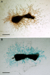
Fig. 1
Fig. 1
β-Galactosidase activity in capillary outgrowth of metatarsals. Cultures of metatarsals of 17-day-old RPTPμ-knock-out/LacZ knock-in fetuses were stained for the endothelial cell marker PECAM (CD31) (a) and LacZ (b). The capillary outgrowth shows a clear blue staining, illustrating the expression of RPTPµ in these cells. Bars represent 200 µm
Fig. 2
Fig. 2
Expression of RPTPµ in bone. Calvariae (a), metatarsals (b) and tibiae (c) of 5-day-old neonatal and tibiae (d) of adult RPTPμ-knock-out/LacZ knock-in mice were stained for β-galactosidase activity using X-gal. Within the calvariae, the cellular network of osteocytes is stained blue. In neonatal metatarsals and tibiae, the osteocytes (arrows) show a deep blue color, while the osteoblasts (arrowheads) are not stained. The adult tibiae also show that the blue staining representing RPTPµ expression is only present in osteocytes (arrows), while lining cells (arrowheads) are negative. The cellular processes (cp) are clearly visible. Bars: a 50 µm; b 100 µm; c, d 25 µm
Fig. 3
Fig. 3
Osteocytes can be generated in vitro. Bone marrow stromal cells (MSC) of RPTPμ-knock-out/LacZ knock-in mice were cultured for 21 days under osteogenic conditions and stained for β-galactosidase activity using X-gal. Within the cultures, blue-stained cells can be observed (a). The number of blue-stained cells is markedly increased when BMPs were added to the medium (b). Close observation shows that within the mineralized (M) nodules osteocytes are present, although the number of blue-stained cells is higher in the non-mineralized areas of the nodules. Histological analysis of the MSC cultures shows that only cells that are embedded within matrix show β-galactosidase activity (c). Some of these cells show cellular processes, which is very characteristic for osteocytes, which can be observed in a larger magnification of the same area (arrowheads d). Bars a, b 250 µm; c, d 25 µm
Fig. 4
Fig. 4
BMPs stimulate, while Noggin inhibits the differentiation of osteocytes. Bone marrow stromal cells (MCS) of RPTPμ-knock-out/LacZ knock-in mice were cultured under osteogenic conditions for 21 days. Thereafter, the cell cultures were processed for β-galactosidase activity analysis by a LacZ enzymatic assay (a) or X-gal staining (b, c). Mineralization of the cultures was examined by alizarin red S staining (b, c). The enzymatic assay shows that BMP-6 clearly stimulates, while Noggin inhibits osteocyte formation. Staining of the cultures with X-gal confirms this observation. The wells stimulated with BMP-6 show more blue staining than the control wells; the wells cultured in the presence of Noggin show hardly any blue staining (b). The same observation can be made from the larger magnification of the wells (c). Alizarin red S staining is also stimulated by BMP-6 and reduced by Noggin, but to a lesser extend (b, c). Bars 200 µm; **p < 0.01; *** p < 0.001
Fig. 5
Fig. 5
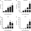
Expression of osteoblast and osteocyte markers. RNA was isolated from RPTPμ-knock-out/LacZ knock-in MSC cultures at days 7, 10, 14, 17 and 21 of differentiation. Expression of osteoblast [alkaline phosphatase (a) and osteocalcin (b)] and osteocyte [LacZ (c) and Sost (d)] markers was analyzed by quantitative RT-PCR with the housekeeping gene β2-microglobulin as internal control. Measurements were performed in triplo, and results are shown as expression relative to day 7 (ac) or day 14 (d), the first day that expression could be shown, calculated using the 2−ΔΔCt method. ***p < 0.001 compared with day 7 (ac) or day 14 (d)
Fig. 6
Fig. 6
Analysis of bone micro-architecture by ex vivo µCT. Bone micro-architecture of RPTPμ-knock-out/LacZ knock-in mice and wild-type FVB mice was analyzed by scanning tibiae of 8-, 20-, 32-, 44- and 56-week-old mice (n = 5–9 per genotype/age group) with a SkyScan 1076 X-ray microtomograph. RPTPμ-knock-out/LacZ knock-in mice show a significantly lower cortical area (a) at 20 weeks in the proximal tibia compared with wild-type mice. Cortical thickness (b) was unaltered and cortical perimeter (c) was slightly lower in RPTPμ-knock-out/LacZ knock-in mice, although this difference was not significant (p = 0.08). RPTPμ-knock-out/LacZ knock-in mice show a slightly lower trabecular bone volume at 20 weeks (d), although this difference was not significant (p = 0.08). Trabecular thickness (e) was unaltered and trabecular connectivity density (f) was significantly lower in RPTPμ-knock-out/LacZ knock-in mice. *p < 0.05

References

    1. Atkins GJ, Findlay DM. Osteocyte regulation of bone mineral: a little give and take. Osteoporos Int. 2012;23:2067–2079. doi: 10.1007/s00198-012-1915-z. - DOI - PubMed
    1. Balemans W, Ebeling M, Patel N, Van Hul E, Olson P, Dioszegi M, Lacza C, Wuyts W, Van Den Ende J, Willems P, Paes-Alves AF, Hill S, Bueno M, Ramos FJ, Tacconi P, Dikkers FG, Stratakis C, Lindpaintner K, Vickery B, Foernzler D, Van Hul W. Increased bone density in sclerosteosis is due to the deficiency of a novel secreted protein (SOST) Hum Mol Genet. 2001;10:537–543. doi: 10.1093/hmg/10.5.537. - DOI - PubMed
    1. Bianchi C, Sellke FW, Del Vecchio RL, Tonks NK, Neel BG. Receptor-type protein-tyrosine phosphatase mu is expressed in specific vascular endothelial beds in vivo. Exp Cell Res. 1999;248:329–338. doi: 10.1006/excr.1999.4428. - DOI - PubMed
    1. Bivi N, Pacheco-Costa R, Brun LR, Murphy TR, Farlow NR, Robling AG, Bellido T, Plotkin LI. Absence of Cx43 selectively from osteocytes enhances responsiveness to mechanical force in mice. J Orthop Res. 2013;31:1075–1081. doi: 10.1002/jor.22341. - DOI - PMC - PubMed
    1. Bonewald LF. The amazing osteocyte. J Bone Miner Res. 2011;26:229–238. doi: 10.1002/jbmr.320. - DOI - PMC - PubMed

Publication types

Substances

LinkOut - more resources